10.2 POJA-L2125+2126+2010+2113+2114+2144
Title: Strata granulosum and corneum of the epidermis
Description:
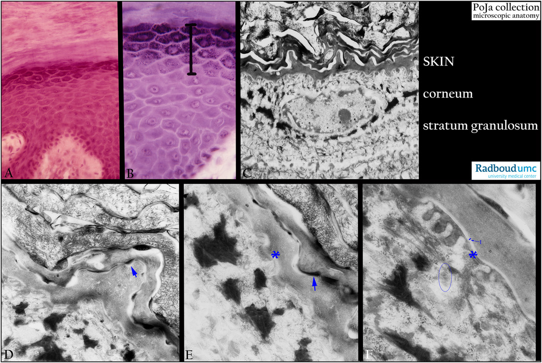
(A): Fingertip, stain hematoxylin-azophloxin, human.
(B): Foot sole, stain hematoxylin-eosin, human. The bar in (B) indicates the stratum granulosum layer that is detailed in (C)
as a transition towards the corneum above the stratum granulosum.
(C-F): Fingertip, electron microscopy, human. Border area between the upper granular cells and the first corneocytes
(stratum corneum, cornified layer).
Electron-dense keratohyalin aggregates (D, F) in the stratum granulosum and the keratinizing fibrillary corneocytes in
the translucent layer (stratum lucidum). (Arrows in D, E) Remnants of desmosomes. (*) Presence of desmosome.
Encircled an intracellular Odland body (lamellar granule) in (F). (1) Points to a white zone, being the cell envelope in (F).
Keywords/Mesh: skin, epidermis, stratum granulosum, granular layer, stratum lucidum (translucent layer), stratum corneum,
cornified layer, desmosome, keratohyalin, histology, electron microscopy, POJA collection

