2.1 POJA-L925
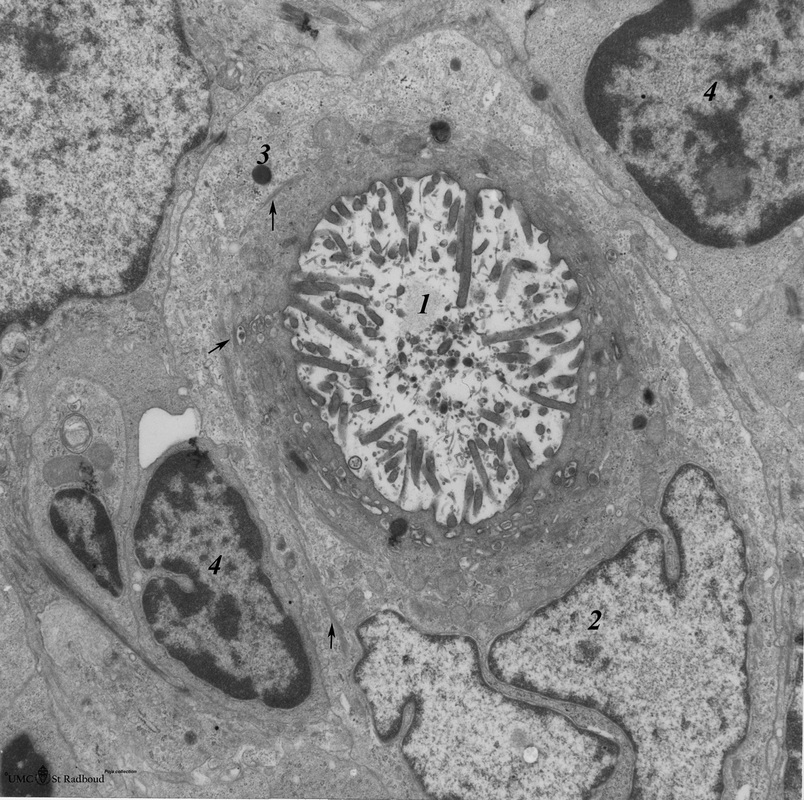
Title: Cystic corpuscle in thymus (mouse, young adult)
Description: Electron microscopy.
This cystic corpuscle is multilayered by specialized epithelioreticular cells and exhibits a lumen (1) which is filled with long microvilli however, curiously derived from a flattened epithelial cell (2).
(3): The outer cells contain few electron-dense keratohyalin granules (3). (→) point to thin bundles of intermediate filaments (keratin filaments).
(4): Thymocytes.
Keywords/Mesh: lymphatic tissue, thymus, cystic corpuscle, Hassall’s corpuscle, thymic corpuscle, histology, electron microscopy, POJA collection

