6.1 POJA-L2954+2675+2676+La0191
Title: Leydig cells in testes
Description:
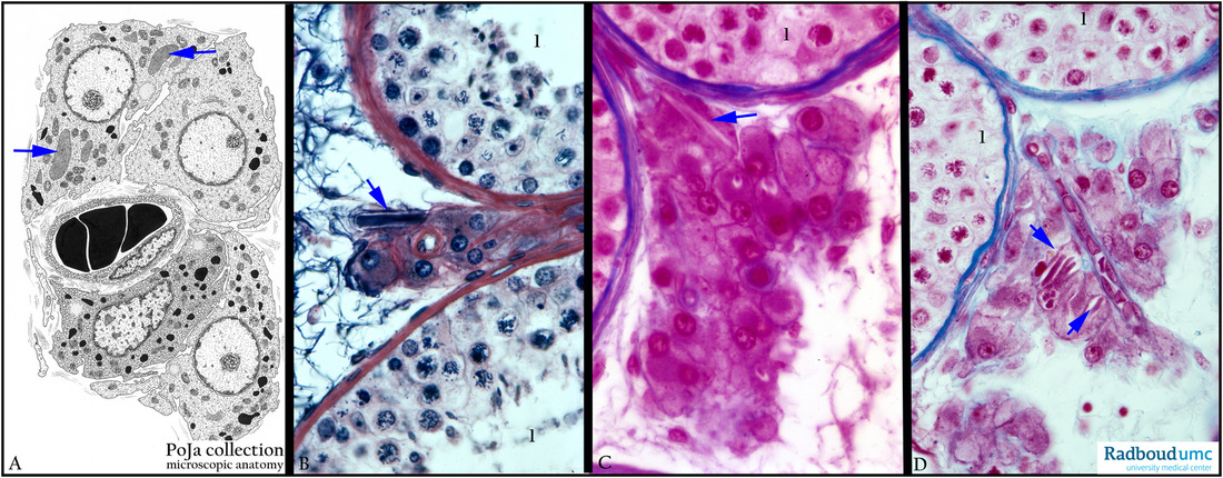
(A) Electron microscopy scheme. (B) Stain iron hematoxylin-eosin, human. (C, D) Stain Azan, human.
The electron micrograph scheme in (A) shows four Leydig cells arranged around a capillary, all located interstitially between seminiferous tubules. Stimulated by the pituitary hormone LH (luteinizing hormone) cholesterol will be converted into pregnenolon leading to the synthesis of testosterone, androstenedion and others. For that purpose the Leydig cells are equipped with ample SER in form of vesicles, membrane-bound lipid droplets, tubular mitochondria, some lipofuscin pigment, and most characteristically the rod shaped crystal-like structures the so-called Reinke crystals, displaying a grid-like organization of proteins. The Reinke crystals are indicated with arrows in
(A-D). Seminiferous tubules (1) are filled with all differentiation stages of the spermatogenesis.
Keywords/Mesh: testis, seminiferous tubule, interstitial Leydig cell, histology, electron microscopy, POJA collection

