
7.1. POJA-L1489+1490+1491+1486Mature cystic teratoma, ovary (human, adult)
7.1. POJA-L1489+1490+1491+1486
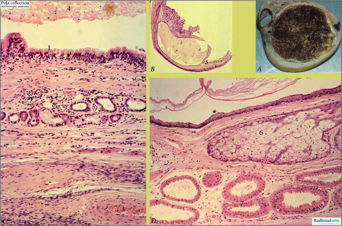
Title: Mature cystic teratoma, ovary (human, adult)
Description: (A) Macroscopy. Stain: (B, C, D) Hematoxylin-eosin.
(A): Macroscopic section of tumor and its dark sebum-like hairy content.
(B): Survey shows cyst surrounded by connective tissue with ectodermal structures (1) including skin and appendages (5).
(C): Lining respiratory epithelium (1) of cavity, fibrous proper lamina with mixed glands (2) and some chronic infiltrate. Blood vessel within fibrocellular tissue (3). Cellular debris (4) in cyst.
(D): Thin keratinized epidermis (*), sebaceous gland (6) and apocrine sweat glands (7). (By courtesy of G. P. Vooijs MD, PhD, former Head of the Department of Pathology, and the Museum of Anatomy and Pathology, Radboud university medical center, Nijmegen, The Netherlands).
Background: Teratomas are mostly benign cystic tumors (hence, the clinical name dermoid cysts) between 5 and 15 cm. Generally the cysts are lined by slightly keratinized squamous epithelium, connective tissue and skin appendages or respiratory epithelium and contain sebaceous material and hairs. Hair shafts protrude from the epidermis and tooth structures and calcified areas are often found within the wall. Frequently fat, bone, cartilage, neural tissue, salivary gland tissue and thyroid tissue are encountered.
Actually teratomas are germ cell neoplasms and consist of displaced ectodermal structures along the line of embryonic fusion resulting in the presence of a mixture of variable tissues. The current parthenogenetic theory suggests origin from a meiotic germ cell. Because the karyotype of benign ovarian teratomas is 46, XX it is therefore suggested that the tumors arise from an ovum after the first meiotic division. Dermoid cysts are sometimes incorporated within the wall of a mucinous cystadenoma. In few cases the dermoids undergo malignant transformation of any of the tissue components e.g. thyroid carcinoma, but most commonly is the occurrence of squamous cell carcinoma.
Keywords/Mesh: female reproductive organs, ovary, cystic teratoma, female genitalia, ovarian neoplasms, dermoid cyst, ovarian tumor, histology, pathology, macroscopy, histology, POJA collection