11.2 POJA-L3256+3273+3229+3453
Title: Myelinated peripheral nerve fibers 7
Description:
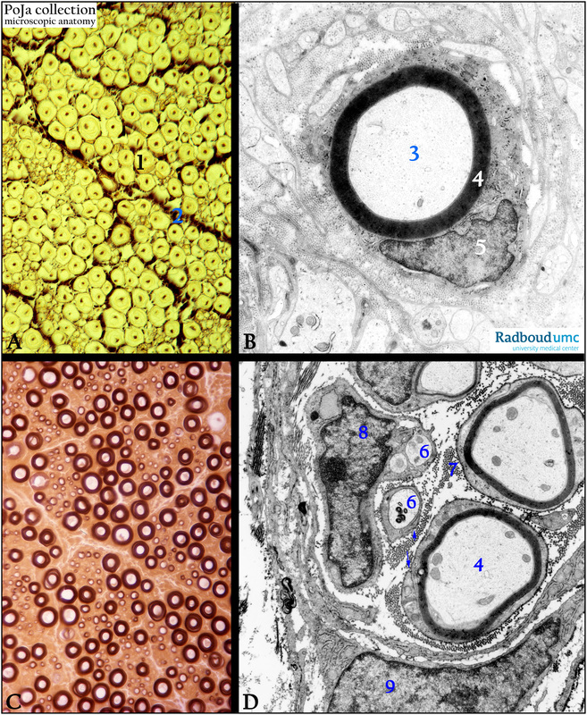
(A): Stain Weigert-osmium, human. (1) Individual myelinated nerve fiber. The axon is visible as a dark “dot”, the surrounding lipid-rich myelin is stained lightly. (2) Perineurium surrounding a whole bundle of nerve fibers.
(B): Electron micrograph, rat. A myelinated (B, 4) nerve fiber (axon, (3) with a cell of Schwann (5).
(C): Stain picroblack blue, human. The myelin of the nerve fibers is stained dark brown, the white central area represents the axon.
(D): Electron micrograph, tannin-fixed/stained, mouse. Equivalent structure to (B, C).
(D, 4) Myelinated axon with neurotubuli and neurofilaments, note the dense-stained fuzzy basal lamina around each Schwann cell. The arrows point to the mesaxon.
(D, 6) Unmyelinated axons enclosed in the wrap of a Schwann cell (D, 8).
(D, 7) Collagen fibers as part of the endoneurium.
(D, 9) Fibroblast with long thin processes as part of the endo- and perineurium. The connective tissue of the perineurium contains several concentric layers of lamellae, between which epithelioid fibroblasts are scattered.
Keywords/Mesh: nervous tissue, axon, peripheral nerve fiber, myelinated nerve fiber, unmyelinated nerve fiber, Schwann cell, endoneurium, histology, electron microscopy, POJA collection

