
12.1.4 POJA-L2570+3575+
2598+2574
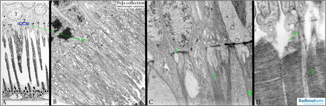
Photoreceptor cells in the retina
12.1.4 POJA-L2570+3575+2598+2574
Title: Photoreceptor cells in the retina
Description:
(A): Deep part of retina, electron microscopy scheme of the outer and inner segment, human.
(1) Nucleus of a cone receptor cell.
(2) Rod cell. Their nuclei are located in the outer nuclear layer. The blue rectangle set represents the desmosomal junctional complexes which all together create the outer limiting membrane.
(3) The thick inner segment of the cone cell contains numerous long and slender mitochondria compared with that of the rod segment.
(4) The outer segment is loaded with parallel packed lamellar discs with the photosensitive transmembrane proteins iodopsin (cones)
and rhodopsin (rods). This segment is connected to the inner segment by a modified cilium.
(5) The surface of the pigmented epithelium is folded into numerous microvilli between which the outer segments are studded.
The dark granules are melanin pigments. Note the distinct basal labyrinth of the cells close to the choriocapillary layer.
(B): Deep part of retina, electron micrograph photoreceptor cells, dog. Only the thin inner segments of the rods are shown here.
(6) Outer limiting membrane as detail of the blue frame in (A). (By courtesy of A. Stadhouders PhD, former Head of the Department of Electron Microscopy, Radboud University, Nijmegen,
The Netherlands).
(C): Deep part of retina, electron micrograph photoreceptor cells, pigeon.
(6) Outer limiting membrane as detail of the blue frame in (A).
(8) Processes of Müller cells (Müller cell fibre baskets) beyond the outer limiting membrane. These long villous processes occupy
the spaces between the inner segments of the photoreceptor cells.
Background: These Müller cells are specialised glia cells responsible for homeostatic and metabolic support of retinal neurons.
They control the composition of extracellular space fluid, regulate the BRB (blood-retina-barrier), and provide trophic and
anti-oxidative support of cones /rods and other neurons.
(D): Deep part of retina, electron micrograph photoreceptor cells, dog. (7) Packed lamellar discs in the outer segments of rods and cones. Arrow points to modified cilium which forms a connection to the inner segment.
Keywords/Mesh: eye, retina, photoreceptor, rod, cone, Müller cell, histology, electron microscopy, POJA collection