
10.3 POJA-L2274+2240+
2088+2241+2037B+2041.
Vater-Pacini corpuscles II
10.3 POJA-L2274+2240+2088+2241+2037B+2041
Title: Vater-Pacini corpuscles II
Description:
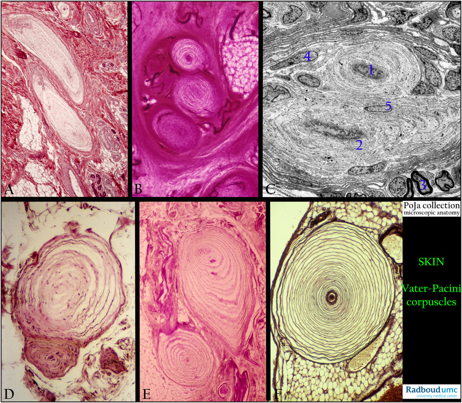
The pacinian corpuscles function as pressure and vibration receptors, and as transducer which responds to mechanical vibrations,
pressure and tensions. The lamellae and the interlamellar space that is filled with liquid, probably amplify any distortion or movement
and thus improve the signal.
(A): Fingertip, stain hematoxylin-azophloxine, human. Note also the presence of fat cells, eccrine sweat glands and their ducts.
(B): Fingertip, Indian ink perfusion-eosin, human. The blood vessels (dark) penetrate up to the periphery of the Pacinian corpuscle,
but not into the center of the corpuscle.
(C): Snout skin, electron microscopy, pig. (1) Inner bulb with afferent axon, rich in mitochondria.
(2) Layers of the perineural sheath.
(3) Myelinated axon.
(4) Fibroblast of the connective tissue sheath.
(5) Schwann cell.
(D): Fingertip, stain hematoxylin-azophloxine, human.
(E): Head skin, stain hematoxylin-eosin, human.
(F): Pancreas, silver stain (Movat), cat. The basal membranes between the perineural cells is visualized by the silver stain.
In the pancreas it provides a mechanism for proprioception, related to the peristaltic movement of the gut.
Keywords/Mesh: skin, Vater-Pacini corpuscle, pacinian corpuscle, sensoric receptor, histology, electron microscopy, POJA collection
Title: Vater-Pacini corpuscles II
Description:
The pacinian corpuscles function as pressure and vibration receptors, and as transducer which responds to mechanical vibrations,
pressure and tensions. The lamellae and the interlamellar space that is filled with liquid, probably amplify any distortion or movement
and thus improve the signal.
(A): Fingertip, stain hematoxylin-azophloxine, human. Note also the presence of fat cells, eccrine sweat glands and their ducts.
(B): Fingertip, Indian ink perfusion-eosin, human. The blood vessels (dark) penetrate up to the periphery of the Pacinian corpuscle,
but not into the center of the corpuscle.
(C): Snout skin, electron microscopy, pig. (1) Inner bulb with afferent axon, rich in mitochondria.
(2) Layers of the perineural sheath.
(3) Myelinated axon.
(4) Fibroblast of the connective tissue sheath.
(5) Schwann cell.
(D): Fingertip, stain hematoxylin-azophloxine, human.
(E): Head skin, stain hematoxylin-eosin, human.
(F): Pancreas, silver stain (Movat), cat. The basal membranes between the perineural cells is visualized by the silver stain.
In the pancreas it provides a mechanism for proprioception, related to the peristaltic movement of the gut.
Keywords/Mesh: skin, Vater-Pacini corpuscle, pacinian corpuscle, sensoric receptor, histology, electron microscopy, POJA collection