
5.4.3 POJA-L2504+2432+
2453+2452+2454+4275+
4274.
Connecting tubule (CNT) (IX) in the human kidney
5.4.3 POJA-L2504+2432+2453+2452+2454+4275+4274
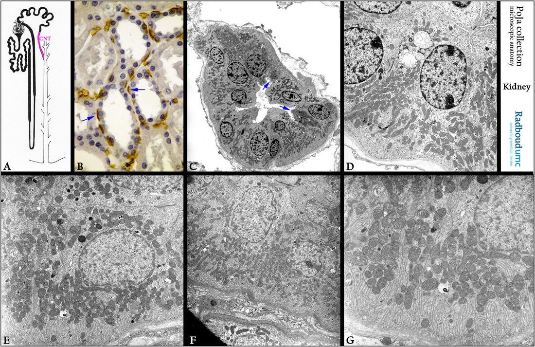
Title: Connecting tubule (CNT) (IX) in the human kidney
Description:
(A): Scheme nephron with CNT in pink color.
(B): Kidney cortex, immunoperoxidase staining with DAB and antibodies against erythrocyte anion exchanger 1 (AE1, AE2, AE3)
or Band 3 protein. Blue arrows indicate a basolateral staining in the intercalated cells of the CNT.
Due to endogenous peroxidase erythrocytes also stained positive.
(C): Electron micrograph of a cross-section through the CNT. Arrows indicate intercalated cells identifiable by their rich content
of apical vesicles and the presence of some small microvilli.
(D): Electron micrograph of a cross-section through a CNT with long small basal folding and mitochondria (near to a glomerulus,
not shown).
(E - G): Electron microscopy, basal regions of various CNTs. Note the characteristic numerous mitochondria accumulated between the basal long and narrow invaginations and folds.
Keywords/Mesh: urinary system, kidney, connecting tubule, CNT, Band 3 protein, histology, electron microscopy, POJA collection