
11.5 POJA-L3015+3058.
Pyramidal cells and neuropil in cerebrum
11.5 POJA-L3015+3058
Title: Pyramidal cells and neuropil in cerebrum
Description:
(A): Stain Kluver-Barrera. The pyramidal neurons are embedded in neuropil, i.e. glia cells and (un)myelinated axons, human.
Capillaries with blue-stained erythrocytes are present.
(B): Electron micrograph of the neuropil of cerebrum, rat. The electron-dense stained rings are myelinated axons. Note also the synapses (green arrows) and presynaptic transmitter vesicles, as well as unmyelinated axons.
(By courtesy of T. Hafmans BSc, Department of Biochemistry, University Medical Centre Radboud University, Nijmegen, The Netherlands).
Keywords/Mesh: nervous tissue, cerebrum, neuron, glia cell, neuropil, myelin, axon, synapse, synaptic vesicle, histology, electron microscopy, POJA collection
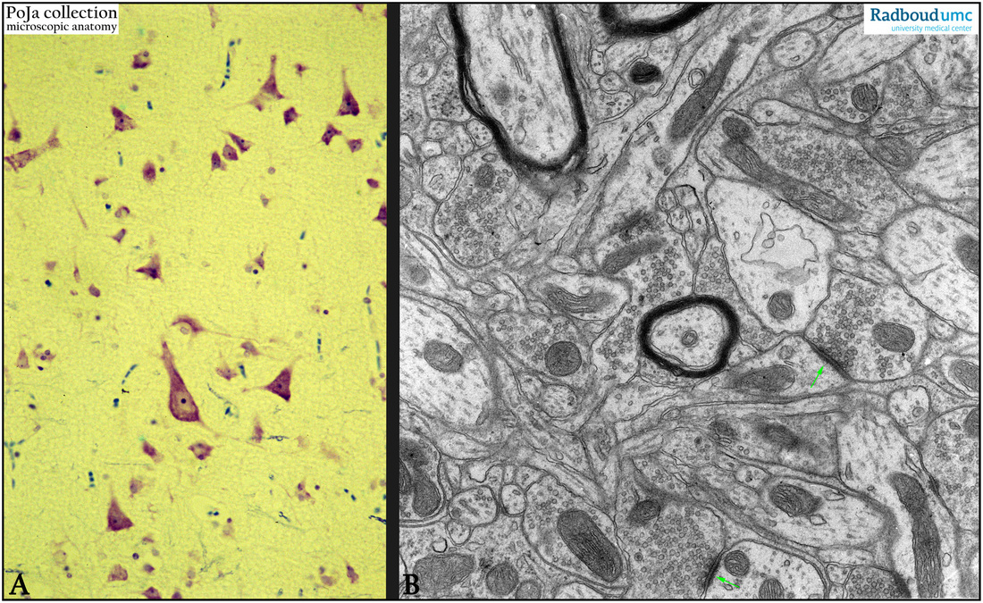