
4.2.1 POJA-L3723. Ruthenium red staining of liver cells (rat)
4.2.1 POJA-L3723
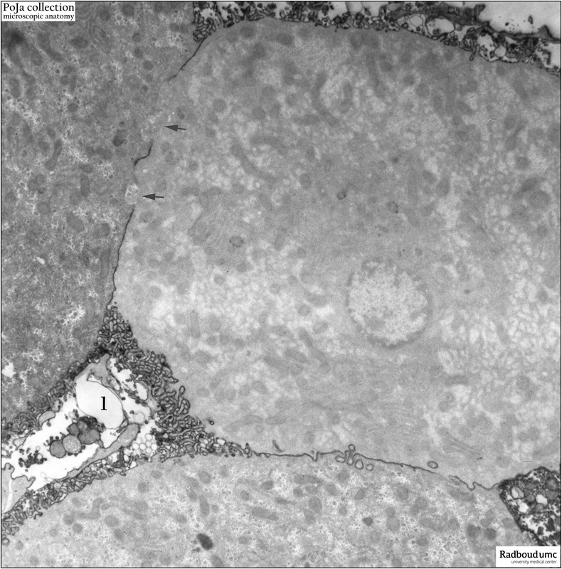
Title: Ruthenium red staining of liver cells (rat)
Description: Electron micrograph of non-contrasted hepatocytes. The ruthenium red stains adjacent liver cell membranes and those bordering the sinusoid (1), their microvilli within the space of Disse. Also endothelial cells are stained. Note however that the bile canaliculi (arrows↘) are impermeable for the substance and remain unstained, as they are sealed off by junctional complexes.
Keywords/Mesh: liver cell, space of Disse, ruthenium red, bile canaliculi, electron microscopy, POJA collection, histology