
5.4.1 POJA-L2469+2473+
2470+2471+2538.
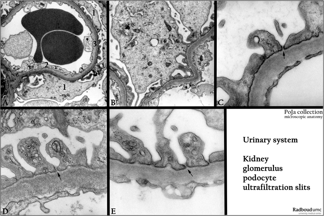
Ultrafiltration slits between pedicles of the podocyte in the glomerulus (XII) of the kidney
5.4.1 POJA-L2469+2473+2470+2471+2538
Title: Ultrafiltration slits between pedicles of the podocyte in the glomerulus (XII) of the kidney
Description:
(A): Glomerulus, electron microscopy, human. Glomerular capillary with apposed podocyte with primary and secondary pedicles or
foot processes (1). (2) Endothelial cell of the capillary. (3) Glomerular basal lamina (GBM).
(B - E): Glomerulus, electron microscopy, human. Details of different podocyte pedicles that create ultrafiltration slits or filtration slit membranes (arrows). The thin filtration slit or gap is estimated between 30 and 60 nm.
Abnormalities in the podocyte slit pattern is often associated with excessive protein loss (nephrotic syndrome).
Keywords/Mesh: urinary system, kidney, glomerulus, capillary, fenestrated endothelium, podocyte, pedicle, ultrafiltration slit, fenestration slit membrane, histology, electron microscopy, POJA collection