
13.1 POJA-L2980+2982+4568+4689 Muscular artery (human)
13.1 POJA-L2980+2982+4568+4689
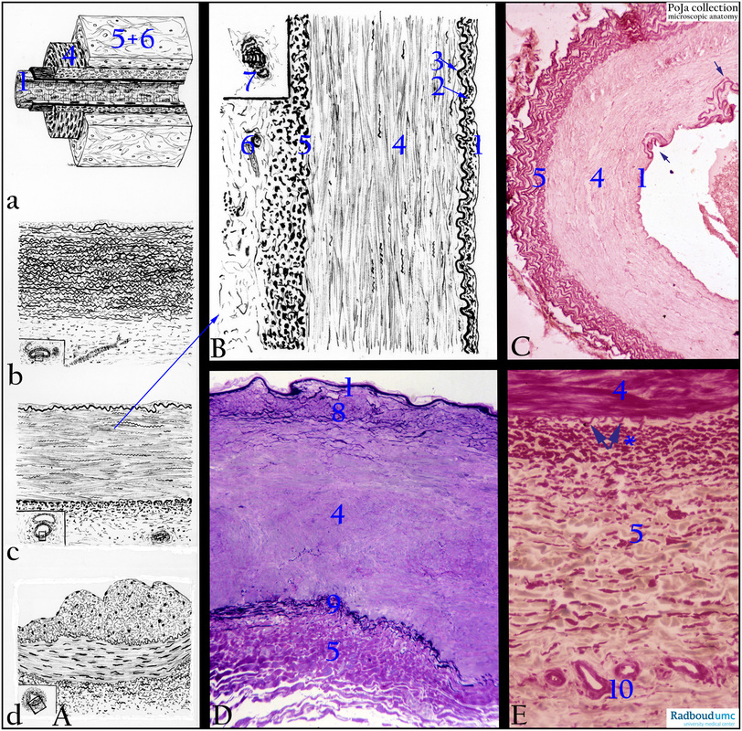
Title: Muscular artery (human)
Description:
(A): Scheme various types of arteries. (A-a): 3D scheme muscular artery. (A-b): Elastic artery. (A-c): Muscular artery, detailed in (B). (A-d): “Sperr”-artery (arterial constrictor) (see 13.1 POJA-L4317+4617+4604+4605+4606 Arterial constrictors). (1) Endothelium. (2) Subendothelial layer (1+2= tunica intima). (3) Internal elastic lamina or I.E.L. (a fenestrated band of elastic fibres at the boundary of intimal and medial tunics). (4) Tunica media (mainly circular smooth muscle cells and supported by a framework of variable numbers of collagen fibres (that limited the distensibility of the arterial wall), extracellular matrix (ECM) and elastic sheaths with irregular perforations. In conducting vessels (elastic arteries), the amount of elastic sheaths is increased and smooth muscle bundles (strain muscles) permeate between the elastic lamellae. In distributing vessels (muscular arteries), the amount of elastic components is reduced and that of smooth muscle fibres increases. (5) External elastic lamina or E.E.L. (fenestrated layer at the boundary of medial and adventitial tunics). (6) Tunica adventitia (loose connective tissue with vasa /nervi vasorum and lymphatics). (7) Vasa vasorum.
(B): Scheme muscular artery (dorsal pedis artery), see (A).
(C): Brachial artery, orcein. Generally mid-sized arteries about 3 mm are distributing arteries and these vessels are characterised by a distinct internal elastic lamina (IEL) between the intima (1) and media (4). A muscular media (4) with finely distributed elastic fibres between the circular orientated smooth muscle cells (SMC), subsequently an external (5) elastic lamina (EEL) as the first lamella followed by numerous short elastic fibres in the adventitia. Arrows point to multiplicated IEL occurring with increasing age.
(D): Popliteal artery (muscle biopsy), toluidine blue-stained 1 micrometer plastic section. In this biopsy the intima is thin and closely followed by the thick IEL (1). In the upper 1/3 of the media (8) increase of elastic fibres, media with SMCs (4) and the boundary with the adventitia (5) is only marked by circular concentrations of elastic fibres (9).
(E): Adventitia femoral artery, resorcin-fuchsin and light green. (4) Media followed by an EEL (external elastic lamina, arrows). (5) Adventitia appears fibroelastic due to a great amount of thick collagen fibres (pale green) and the dense pink-stained elastic fibres (*) with the highest concentration near the EEL. The smaller the vessel the lesser the amount of elastic material. (10) Vasa vasorum.
Keywords/Mesh: cardiovascular system, vascularisation, blood vessel, muscular artery, elastin, histology, POJA collection