5.4.1 POJA-L2474+2369+2540
Title: Mesangium cells in renal glomerulus (XV) of the human kidney
Description:
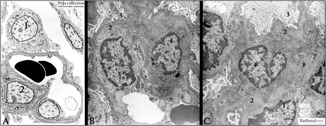
(A): Glomerulus, electron microscopy scheme.
(1) Podocyte.
(2) Mesangium cell.
(B, C): Electron microscopy of human mesangium cells.
(1) Endothelial cell nucleus.
(2) Basal lamina.
(3) Pedicles of a podocyte.
(4) Mesangium cell.
Note the impressively developed basal laminae bordering the extensive mesangial matrix from the podocytes.
Keywords/Mesh: urinary system, kidney, glomerulus, mesangium cell, mesangium, podocyte, histology, electron microscopy, POJA collection

