
2.2 POJA–L1010. Phagocytosis in splenic red pulp (mouse)
2.2 POJA–L1010
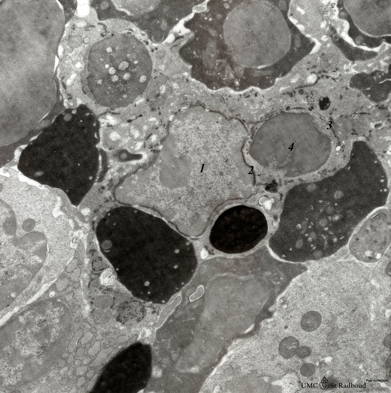
Title: Phagocytosis in splenic red pulp (mouse)
Description: Electron microscopy. Stain: peroxidase reaction with diaminobenzidin staining.
A diversity of red blood cells in the red pulp can be discerned due to the DAB staining of hemoglobin by oxidized benzidin (dark and light staining).
The macrophage (1) does show peroxidase activity along the nuclear membrane (2) and in profiles of the endoplasmic reticulum (3) and in few small lysosomes. Note also a large negative-stained inclusion (4) in its cytoplasm as a remnant of a phagocytosed old erythrocyte. Nucleated erythocytes stained electron-grey to -dark depending on the amount of hemoglobin. Even reticulocytes wih reduced amounts are still stained, while all other non-erythrocytic cells are negative with the exception of a macrophage (or phagocyte) interspersed between all these cells.
Keywords/Mesh: lymphatic tissue, spleen, phagocytosis, histology, electron microscopy, POJA collection