
16.1.2 POJA-L7154 Macroscopy of the femur bone
16.1.2 POJA-L7154 Macroscopy of the femur bone
(By courtesy of J. Kooloos PhD and L. Boer PhD Department Medical Imaging, Anatomy, Museum for Anatomy and Pathology, Radboud university medical center, Nijmegen The Netherlands)
16.1.2 POJA-L7154 Macroscopy of the femur bone
Title: Macroscopy of the femur bone
Description:
Proximal end of right femur.
(1): Head,
(2): Fovea,
(3): Neck,
(4): Greater trochanter,
(5): Lesser trochanter,
(6): Shaft (diaphysis),
(7): The remains of the epiphysial line.
(8): Principal compressive stress:
The stress lines are drawn according to the publication:
(Bio-inspired method based on bone architecture to optimize the structure of mechanical workspieces. C. Audibert, J. Chaves-Jacob, J-m. Linares, Q-A. Lopez in Materials and Design October 2018, https://doi.org/10.1016/j.matdes.2018.10.013
The outer part of the bone is solid and is composed of compact or dense bone. It is distinct in the diaphysis (6), which encloses bone marrow cavity.
The interior shows a spongy configuration the so-called spongy or cancellous bone due to bony trabeculae with a wide variation in thickness, orientation (coloured lines) and spacing between the trabeculae. Between the trabeculae haematopoieteic bone marrow (stem) cells are located.
The 3D orientation of the trabeculae is correlated to the magnitude and direction of forces acting on the head of the femur. In cancellous bone the density of bone is reduced and in this way the ends of long bones are allowed to be compressed as the result of stresses applied to the bone.
There is a characteristic arrangement of the trabeculae i.e. one side of the bone bears the pression and the other side withstands compression. The cancellous bone appears to respond in the same way to the applied forces as cortical bone does. The result is a relation between the trabecular microstructure in biologically mediated adaptation to the mechanical environment. Mechanically, the upper femur consists of two separate bones: the combined femoral head and neck and the femoral shaft. Both bones consist of a mixture of two different types: cancellous bone and cortical bone and are subjected to two different forces: those of weight bearing and movement and those of bone modelling. (The calcar femorale: A new perspective. A. Hammer J.Orthopaedic Surgery https://doi.org/10.1177/2309499019848778)
See also:
Key words/Mesh: locomotor system, bone, compact bone, spongy bone, femur, diaphysis, epiphysis, trabecula, endochondral ossification, macroscopy, histology, POJA collection
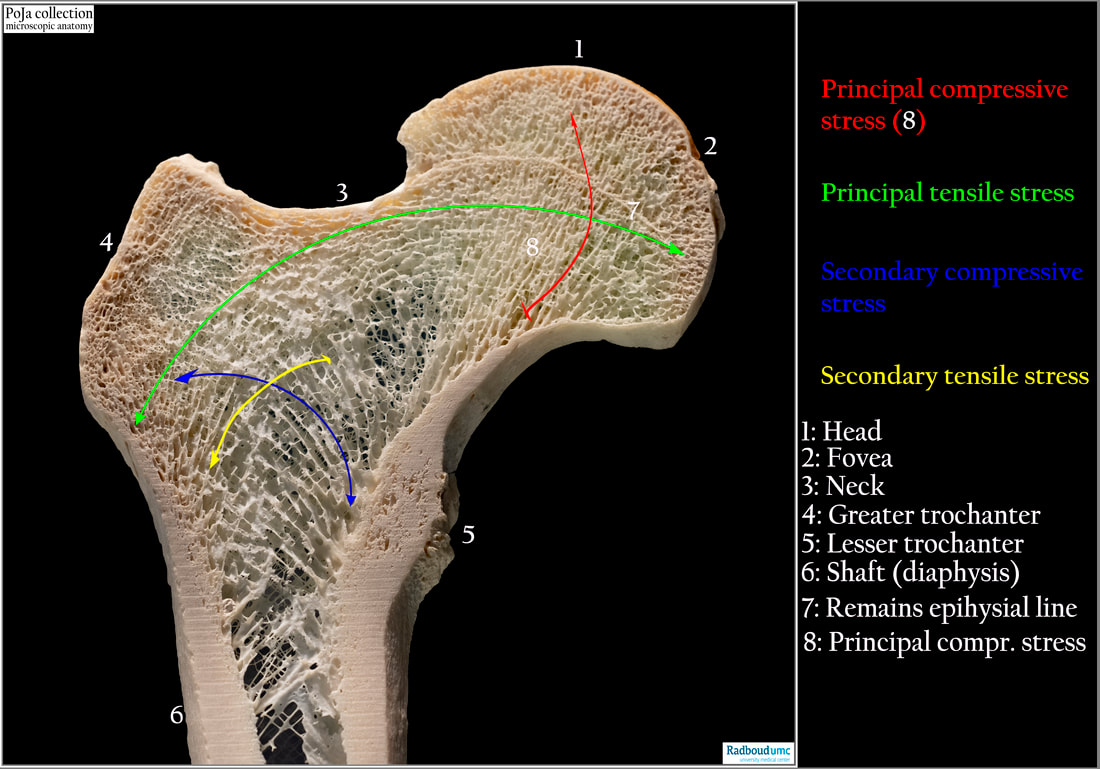
Title: Macroscopy of the femur bone
Description:
Proximal end of right femur.
(1): Head,
(2): Fovea,
(3): Neck,
(4): Greater trochanter,
(5): Lesser trochanter,
(6): Shaft (diaphysis),
(7): The remains of the epiphysial line.
(8): Principal compressive stress:
- Red line segment: with orientated trabeculae perpendicular to the epihysial line.
- Blue line segment: secondary compressive stress.
- Green line segment: principal tensile stress, from area below 7 to the end of the greater trochanter.
- Yellow line segment: from top of the diaphysis to the middle of the bone marrow cavity (The basis of Wolff's trajectorial theory).
The stress lines are drawn according to the publication:
(Bio-inspired method based on bone architecture to optimize the structure of mechanical workspieces. C. Audibert, J. Chaves-Jacob, J-m. Linares, Q-A. Lopez in Materials and Design October 2018, https://doi.org/10.1016/j.matdes.2018.10.013
The outer part of the bone is solid and is composed of compact or dense bone. It is distinct in the diaphysis (6), which encloses bone marrow cavity.
The interior shows a spongy configuration the so-called spongy or cancellous bone due to bony trabeculae with a wide variation in thickness, orientation (coloured lines) and spacing between the trabeculae. Between the trabeculae haematopoieteic bone marrow (stem) cells are located.
The 3D orientation of the trabeculae is correlated to the magnitude and direction of forces acting on the head of the femur. In cancellous bone the density of bone is reduced and in this way the ends of long bones are allowed to be compressed as the result of stresses applied to the bone.
There is a characteristic arrangement of the trabeculae i.e. one side of the bone bears the pression and the other side withstands compression. The cancellous bone appears to respond in the same way to the applied forces as cortical bone does. The result is a relation between the trabecular microstructure in biologically mediated adaptation to the mechanical environment. Mechanically, the upper femur consists of two separate bones: the combined femoral head and neck and the femoral shaft. Both bones consist of a mixture of two different types: cancellous bone and cortical bone and are subjected to two different forces: those of weight bearing and movement and those of bone modelling. (The calcar femorale: A new perspective. A. Hammer J.Orthopaedic Surgery https://doi.org/10.1177/2309499019848778)
See also:
- 16.1.2 POJA-L7112+7074+7117 Lamellar bone (or secondary bone)
- 16.1.3 POJA-L7114+7112+7093+7092 Remodeling of compact bone (cortical bone) 5
- 16.1.3 POJA-L7147+7111+7110 Endochondral ossification in foetus 6
- 16.1.3. POJA-L7116+7115+7123 Endochondral ossification in foetus 7
Key words/Mesh: locomotor system, bone, compact bone, spongy bone, femur, diaphysis, epiphysis, trabecula, endochondral ossification, macroscopy, histology, POJA collection