5.4.3 POJA-La0356+L2374+2390+2391+La0072+2414
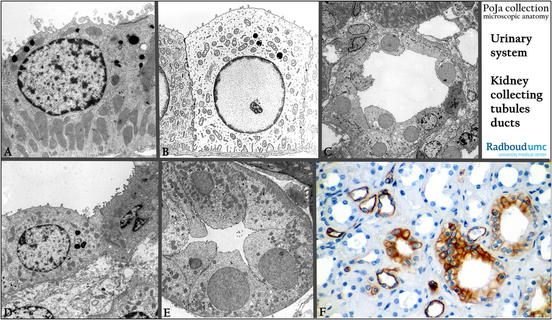
Title: Collecting tubules and ducts (XXII) in rat kidney
Description:
(A): Electron micrograph of mTAL, medullary thick limb of Henle. Mitochondria are arranged between the basal invaginations;
a limited number of short microvilli, and scarce number of lysosomes.
(B): Electron microscope scheme (human) of a cell of the CD, collecting duct. This cell hardly contains basal invaginations,
no microvilli,
(C): Electron micrograph of a cross-section through a outer medullary collecting duct (OMCD) in the inner stripe. (perfusion fixation).
(D): Electron microscopy, similar as (C) showing light and dark intercalated cells (IC). Note that the epithelium in this duct is rather low
to cuboidal.
(E): Electron micrograph of an IMCD, inner medullary collecting duct approximate to the papillary duct.
Note that the epithelial cells are high to columnar.
(F): Immunoperoxidase staining with DAB and monoclonal antibodies against cytokeratin 7 (OVTL12/30).
Note that the mTAL tubules are negative, but the OMCD (outer medullary collecting duct) and DTL (descending thin limb) are positive.
The OVTL12/30-CK7 has been reported negative for proximal convoluted tubules, but Henle’s loops and distal convoluted tubules and collecting ducts are positive.
Keywords/Mesh: urinary system, kidney, cytokeratin 7, collecting tubules, collecting ducts, histology, electron microscopy, POJA collection

