
12.1.5 POJA-L3562+2534+
2591
Lens, iris and ciliary body of the eye
12.1.5 POJA-L3562+2534+2591
Title: Lens, iris and ciliary body of the eye
Description:
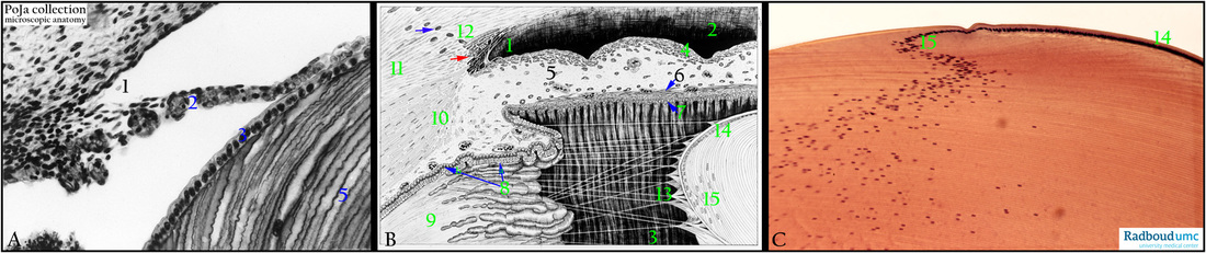
(A): Stain Haematoxylin-eosin (black and white print), embryo human. (1) Corneal-irideal angle. (2) Future iris with capillaries. (3) Anterior lens epithelium. (5) Elongated lens cells.
(B): Scheme, adult human.
- Corneal-irideal angle (1) with anterior chamber (2) and posterior chamber (3).
- Iris (4) with anterior surface without epithelial lining. Loose connective tissue stroma (5) with melanocytes and myoepithelial cells
(dilator pupillae muscle, 6, zoom-in!) close to posterior surface lined by a layer of pigmented epithelial cells (7).
- Ciliary body (8) with ciliary processes (pars plicata) and dual epithelial layer of nonpigmented cells (arrow 8) (faces the posterior chamber and pigmented cells (arrow 8b) (faces the stroma of the ciliary processes).
- Pars plana (9) of the ciliary body.
- Ciliary muscle (10) close to the sclera (11).
- Limbal area (12) with canal of Schlemm (blue arrow) and trabecular meshwork red arrow) of the corneal-irideal angle.
- Suspensory ligament or zonular fibres (13) (or zonules of Zinn, Zinn’s membrane) arise from the pars plana ( the shorters fibres from pars plicata, the longer ones from the pars plana) and form a tent-like structure upon attaching to the lens capsule.
They are produced by the non-pigmented epithelial cells of the ciliary body and contain a.o. collagen type IV as well as fibrillin.
- Anterior lens epithelium (14).
- Equatorial zone (15) with nucleated lens fibres.
(C): Stain Haematoxylin-eosin, pig. Anterior lens epithelium (14). Equatorial zone (15) and elongated nucleated lens cells (=lens fibres).
Keywords/Mesh: eye, lens, ciliary body, iris, zonular fibre, histology, POJA collection
Title: Lens, iris and ciliary body of the eye
Description:
(A): Stain Haematoxylin-eosin (black and white print), embryo human. (1) Corneal-irideal angle. (2) Future iris with capillaries. (3) Anterior lens epithelium. (5) Elongated lens cells.
(B): Scheme, adult human.
- Corneal-irideal angle (1) with anterior chamber (2) and posterior chamber (3).
- Iris (4) with anterior surface without epithelial lining. Loose connective tissue stroma (5) with melanocytes and myoepithelial cells
(dilator pupillae muscle, 6, zoom-in!) close to posterior surface lined by a layer of pigmented epithelial cells (7).
- Ciliary body (8) with ciliary processes (pars plicata) and dual epithelial layer of nonpigmented cells (arrow 8) (faces the posterior chamber and pigmented cells (arrow 8b) (faces the stroma of the ciliary processes).
- Pars plana (9) of the ciliary body.
- Ciliary muscle (10) close to the sclera (11).
- Limbal area (12) with canal of Schlemm (blue arrow) and trabecular meshwork red arrow) of the corneal-irideal angle.
- Suspensory ligament or zonular fibres (13) (or zonules of Zinn, Zinn’s membrane) arise from the pars plana ( the shorters fibres from pars plicata, the longer ones from the pars plana) and form a tent-like structure upon attaching to the lens capsule.
They are produced by the non-pigmented epithelial cells of the ciliary body and contain a.o. collagen type IV as well as fibrillin.
- Anterior lens epithelium (14).
- Equatorial zone (15) with nucleated lens fibres.
(C): Stain Haematoxylin-eosin, pig. Anterior lens epithelium (14). Equatorial zone (15) and elongated nucleated lens cells (=lens fibres).
Keywords/Mesh: eye, lens, ciliary body, iris, zonular fibre, histology, POJA collection