16.1.3 POJA-L7090+7076 Bone: osteons in polarisation microscope
16.1.3 POJA-L7090+7076 Bone: osteons in polarisation microscope
Title: Bone: osteons in polarisation microscope
Description:
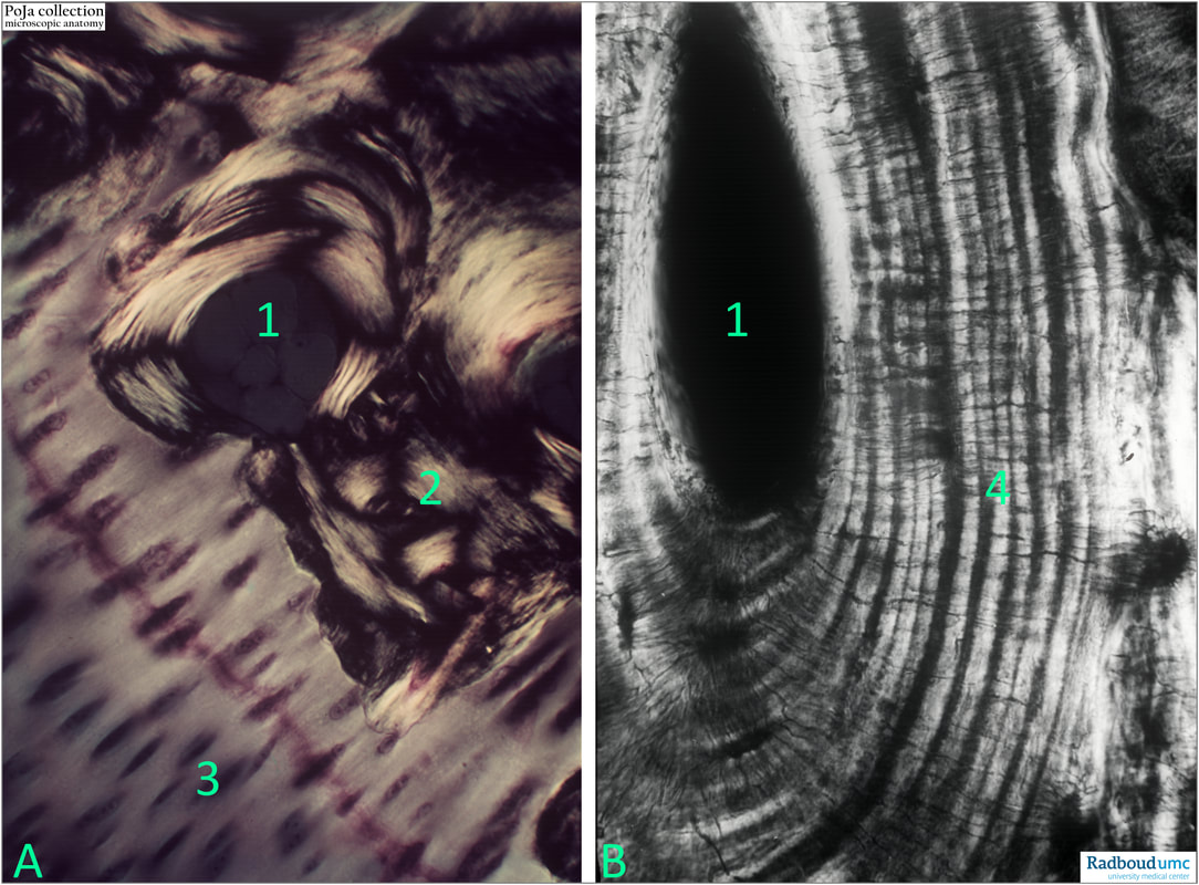
(A+B): Human bone visualised with polarisation microscopy.
(1): Haversian canal (dark area) of an osteon.
(2): Interstitial bone lamellae.
(3): Light collagen fibres part of Sharpey’s fibres. Dark oblong spots are non-birefringent structures with the bundle collagen fibres.
(4): Alternating light and dark lamellae.
Background:
Collagen fibres display optical anisotropy in polarised light. When collagen fibres are transversely aligned to the direction of the light propagation, there is a change in the refraction of light that results in a maximum brightness. When collagen fibres align along the axis of light propagation, no refraction occurs and the result is a dark specimen. Collagen fibres oriented in other directions show no or intermediate brightness.
The Haversian channels are formed by concentric layers called lamellae, which are approximately 50 µm in diameter. Each Haversian canal generally contains one or two capillaries and many nerve fibres. The canals are in fact communication channels with the osteocytes that are positioned in lacunae. Haversian canals are contained within osteons, which are typically arranged along the long axis of the bone in parallel to the surface. The canals and the surrounding lamellae (8-15) form the functional unit, called a Haversian system, or osteon.
Reference:
Circularly polarized light standards for investigations of collagen fiber orientation in bone. Timothy G. Bromage, Haviva M. Goldman, Shannon C. McFarlin, Johanna Warshaw, Alan Boyde, Christopher M. Riggs. https://doi.org/10.1002/ar.b.10031
See also:
Keywords/Mesh: locomotor system, bone, compact bone, osteon, Haversian canal, birefringe, collagen, Malthese Cross effect, polarisation, histology, POJA collection
Title: Bone: osteons in polarisation microscope
Description:
(A+B): Human bone visualised with polarisation microscopy.
(1): Haversian canal (dark area) of an osteon.
(2): Interstitial bone lamellae.
(3): Light collagen fibres part of Sharpey’s fibres. Dark oblong spots are non-birefringent structures with the bundle collagen fibres.
(4): Alternating light and dark lamellae.
Background:
Collagen fibres display optical anisotropy in polarised light. When collagen fibres are transversely aligned to the direction of the light propagation, there is a change in the refraction of light that results in a maximum brightness. When collagen fibres align along the axis of light propagation, no refraction occurs and the result is a dark specimen. Collagen fibres oriented in other directions show no or intermediate brightness.
The Haversian channels are formed by concentric layers called lamellae, which are approximately 50 µm in diameter. Each Haversian canal generally contains one or two capillaries and many nerve fibres. The canals are in fact communication channels with the osteocytes that are positioned in lacunae. Haversian canals are contained within osteons, which are typically arranged along the long axis of the bone in parallel to the surface. The canals and the surrounding lamellae (8-15) form the functional unit, called a Haversian system, or osteon.
Reference:
Circularly polarized light standards for investigations of collagen fiber orientation in bone. Timothy G. Bromage, Haviva M. Goldman, Shannon C. McFarlin, Johanna Warshaw, Alan Boyde, Christopher M. Riggs. https://doi.org/10.1002/ar.b.10031
See also:
- 16.1.3 POJA-L7079+7080+7082+7081 Compact bone (cortical bone) with osteons
- 16.0 POJA-L7087+7088 Partially burned human bone specimen
Keywords/Mesh: locomotor system, bone, compact bone, osteon, Haversian canal, birefringe, collagen, Malthese Cross effect, polarisation, histology, POJA collection

