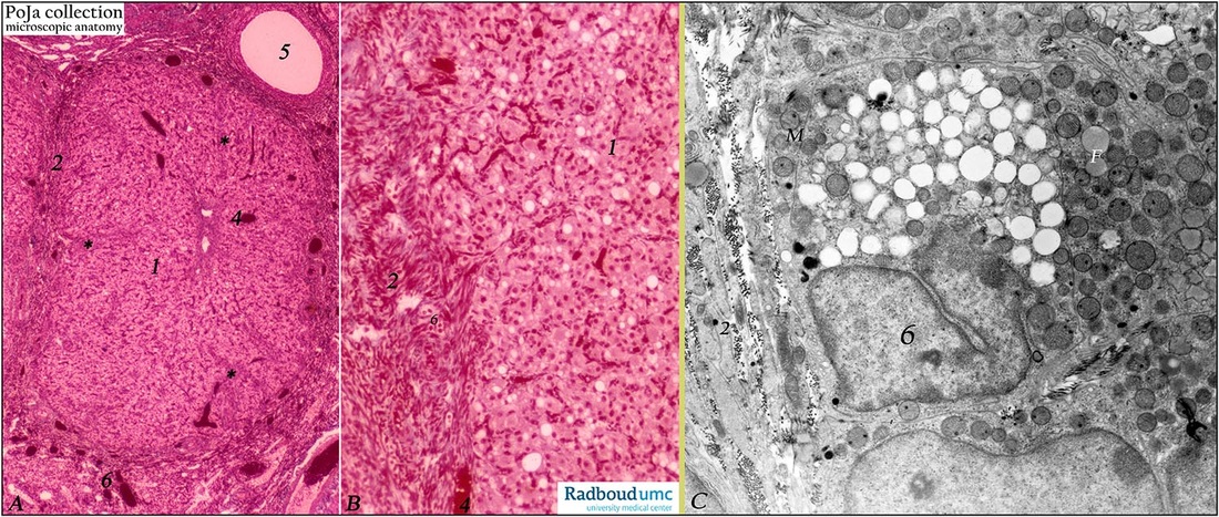
7.1 POJA-L1211+L1236+L1241
Title: Corpus luteum in ovary (rabbit)
Description: (A, B) Stain: Azan. (C) Electron microscopy.
(A): Survey: after ovulation the lining stratum granulosum of the empty follicle collapses, becomes folded. Under influence of luteinizing hormone granulosa cells increase in size (1) and transform into granulosa lutein cells. Accompanying external theca cells provide stroma and septa (2) of the enlarging corpus luteum. The latter possesses a characteristic folded wall (*) supplied by numerous blood vessels (A, B, 4) and supportive septa. Note stroma (6) with blood vessels around corpus luteum and antral follicle (5).
(B): Numerous spindle-shaped or elongated endothelial cells occur between predominantly granulosa lutein cells (1). Septa (2) and stroma with few small theca lutein cells (6).
(C): Ultra structure of granulosa lutein cell (6) with horse shoe-shaped nucleus, tubular mitochondria (m). Note numerous electron-light and -grey lipid globules (F). Elongated stroma cell projections (2); in between them cross-sectioned bundles collagen fibers.
Background: Under influence of the luteinizing hormone (LH) the luteinization process starts i.e. differentiation of granulosa cells into granulosa lutein cells while they produce a yellow carotenoid pigment (lutein) as well as progesterone/estrogen (in response to follicle-stimulating hormone FSH and LH stimulation). Hence the name corpus luteum due to its yellowish color. The basement membrane of the collapsed follicle is broken down and internal theca cells invade into the cellular mass together with capillaries and stromal cells. Theca interna cells differentiate into theca lutein cells (or paralutein cells) and produce androstenedione and progesterone (in response to LH stimulation). Together with predominantly granulosa lutein cells they form epithelial-like clusters and sheets of round/ polygonal lutein cells with lipid droplets (cave hormone production) that comprise the parenchyma of a corpus luteum. Androstenedione is provided by theca lutein cells to the granulosa lutein cells to produce estradiol in the latter cells.
Keywords/Mesh: female reproductive organs, ovary, corpus luteum, luteal cells, theca cells, female genitalia, granulosa cells, histology, POJA collection

