4.1.1 POJA-L4004+4009++4008+4007+4001+4006+4005+4003
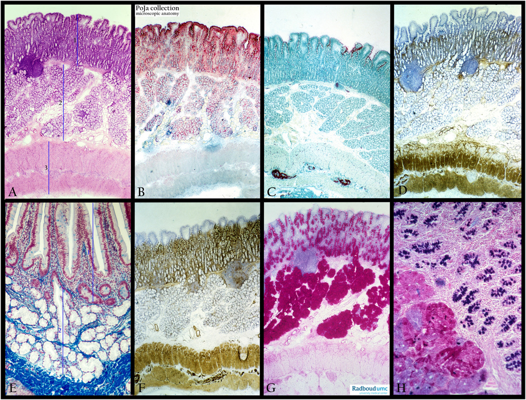
Title: Histochemical profile of duodenum (pig, human)
Description: Stain: (A) Hematoxylin-eosin. (B) ZFFA. (C) AFFA. ( D) AMPase. (E) Azan, human. (F) ATPase. (G) PAS. (H) Alcian blue-PAS.
(A, 1): Comprises the mucosa layer that consists of an epithelium lining that is folded into villi and crypts and is supported by a lamina propria loose connective tissue layer that ends on a small layer of muscle cells, the so called lamina muscularis mucosae. The same layer, in detail is seen in (E, 1).
(A, 2): Is the submucosa zone of connective tissue but filled up with the Lieberkühn crypts and Brunner glands. Identical layer is presented in detail in (E, 2). Note that the epithelium lining also contains mucus producing goblet cells (bluish stained).
Also notice that the white Brunner glands end up in the darker stained crypt epithelium.
(A, 3): Tunica muscularis consists of an inner circular muscle layer and an outer longitudinal muscle layer. Both the glandular cells and the goblet cells are visualized intensively with the PAS (G) and the Alcian blue-PAS (H) stain.
The AMpase and ATPase are strongly expressed in the Lieberkühn crypts and the muscle fibers but much less in the surface epithelium and the Brunner glands.
Keywords/Mesh: duodenum, histochemistry, Brunner glands, Lieberkühn crypts, PAS, histology, POJA collection

