11.2.1 POJA-L3311+3314+3276+3277+3310
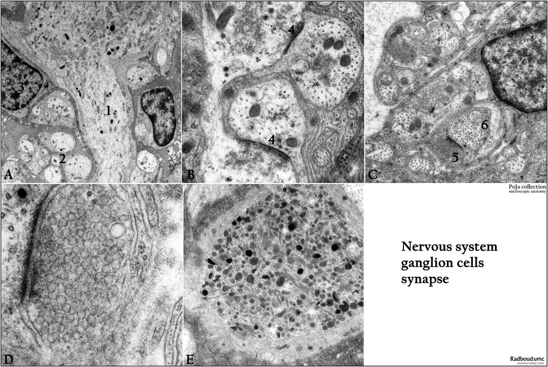
Title: Sympathetic trunk ganglion cells and synapses
Description:
Electron micrographs.
(A): Ganglion cell with axon beginning at the axon hillock (1), rabbit. (2) Additional nonmyelinated axons. (3) Nucleus of
an amphicyte.
(B, D): Two axo-axonal synapses (4) with pre- (thin) and postsynaptic plasmalemmas (thick) and a synaptic cleft between them, rabbit. The presynaptic vesicles with transmitter substances are detailed in (D), rat.
(C): Synapses: (5) Presynaptic end terminal with transmitter vesicles. (6) Postsynaptic part of a nerve cell. Note the thickening of the plasmalemmas and the cleft between the two, rat.
(E): The cytoplasmic part of a myelophage with numerous different types of young and old lysosomes, rabbit These infiltrating macrophages are very active in phagocytosis and removal of degenerated debris between and within nerve fibers hence, the name myelophage.
Keywords/Mesh: nervous system, ganglion cell, synapse, myelophage, histology, electron microscopy, POJA collection

