4.3.1 POJA-L2928+4202
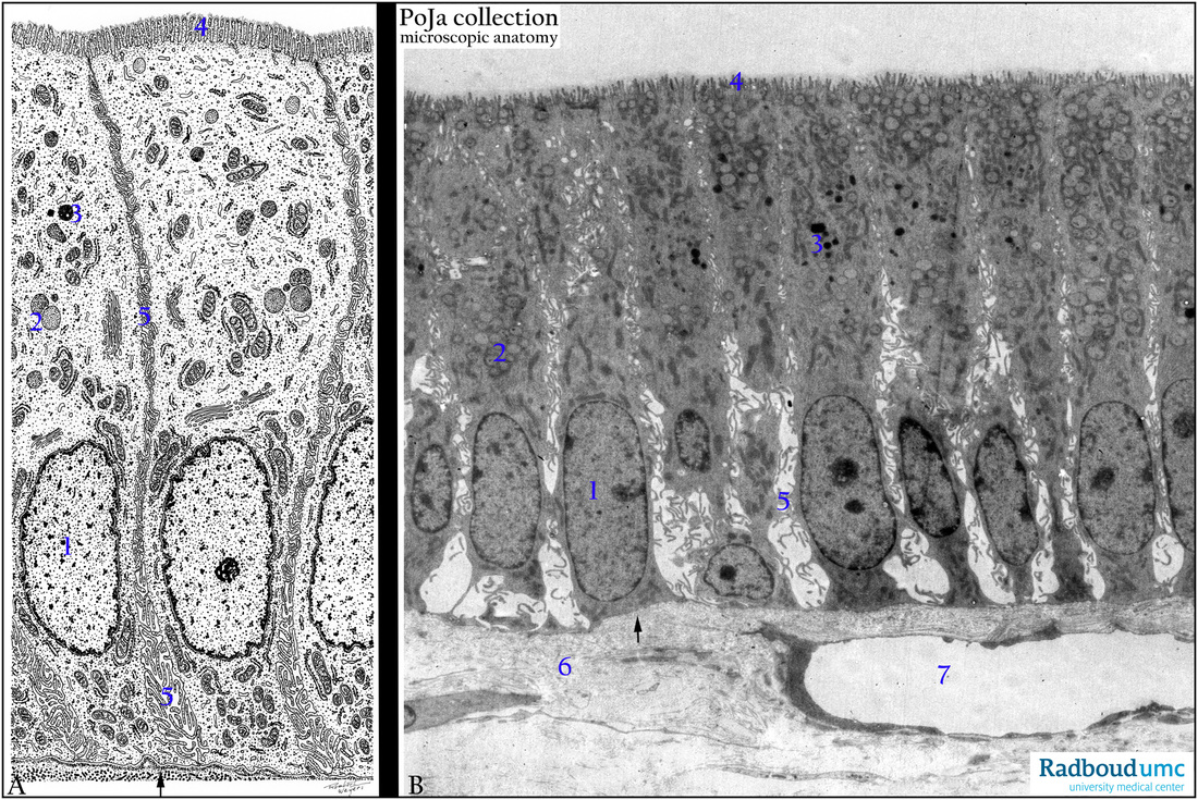
Title: Gallbladder epithelium scheme and electron microscopy (human, monkey)
Description: (A) Scheme and (B) electron micrograph of gallbladder epithelium.
The columnar epithelial cells (1) contain microvilli or brush border (4), electron-grey secretion granules (2) with a mucus content to protect the outer cell surface.
(3) Darkly stained lysosomes.
(5) Intercellular spaces used to transport water from the gall via / through the cell to the vessels (7) in order to concentrate the gall liquid. (6) Loosely arranged lamina propria.
(↘, arrows) indicate the basal lamina.
Note that the cells contain large numbers of elongated mitochondria in order to provide the energy required for the transport and concentration process.
Keywords/Mesh: gallbladder, water transport, bile, epithelium, electron microscopy, histology, POJA collection

