
13.1 POJA-L4653+4654 Fenestrated capillaries and sinusoids
13.1 POJA-L4653+4654
Title: Fenestrated capillaries and sinusoids
Description:
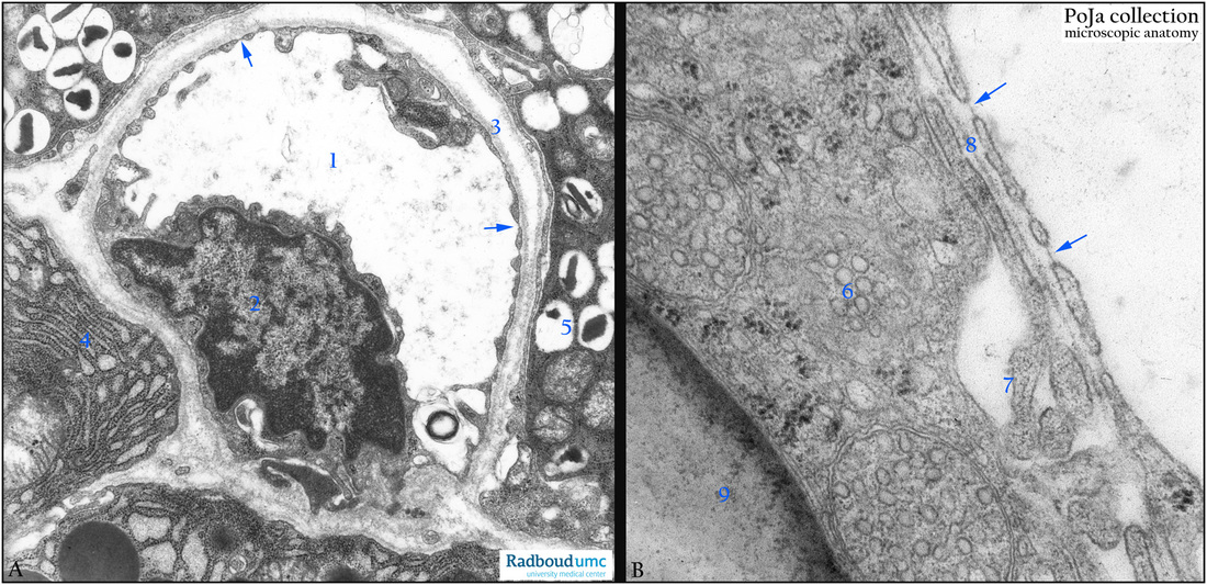
(A): Electron micrograph of fenestrated capillary in pancreas, dog. (1) Lumen capillary. (2) Nucleus endothelial cell. (3) Lamina basalis. (4) Exocrine pancreas cell with extended RER profiles. (5) Endocrine pancreas insulin producing cell. Arrows point to the fenestrae of the endothelial cell.
(B): Electron micrograph of sinusoid in medulla of adrenal gland, rat. Arrows point to the gaps with diaphragm in the lining endothelium of the sinusoid. Lipid droplet (9) in cell of the fasciculata zone and tubular mitochondria (6) both characteristic for hormone producing cells. (7) Intercellular space with microvilli-like extensions. (8) Discontinuous basal lamina.
Background:
1. Capillaries with fenestrated endothelium and an uninterrupted basal lamina are most frequently found in gastrointestinal mucosa, endocrine tissue and kidney. These fenestrae are warrant for an intensive exchange between capillary and surrounding cells. Fenestrae in the endothelium range generally from 20-100 nm (average about 70 nm) in diameter and limited by a thin diaphragm (4 nm thick) with a central opening of 5 nm diameter. The diaphragms are negative charged and mucin-dependent. However, fenestrae without diaphragm are also observed e.g. kidney.
2. Discontinuous capillary or sinusoids are characterised by incomplete basal lamina and gaps (larger than fenestrae) in endothelium (liver, spleen, adrenal gland). They are endothelial-lined channels with a larger diameter than capillaries with discontinuous or absence of basement membranes.
Keywords/Mesh: cardiovascular system, vascularisation, capillary , fenestra, fenestrated endothelium, basal lamina, discontinuous capillary, sinusoid, histology,, electron microscopy, POJA collection
Title: Fenestrated capillaries and sinusoids
Description:
(A): Electron micrograph of fenestrated capillary in pancreas, dog. (1) Lumen capillary. (2) Nucleus endothelial cell. (3) Lamina basalis. (4) Exocrine pancreas cell with extended RER profiles. (5) Endocrine pancreas insulin producing cell. Arrows point to the fenestrae of the endothelial cell.
(B): Electron micrograph of sinusoid in medulla of adrenal gland, rat. Arrows point to the gaps with diaphragm in the lining endothelium of the sinusoid. Lipid droplet (9) in cell of the fasciculata zone and tubular mitochondria (6) both characteristic for hormone producing cells. (7) Intercellular space with microvilli-like extensions. (8) Discontinuous basal lamina.
Background:
1. Capillaries with fenestrated endothelium and an uninterrupted basal lamina are most frequently found in gastrointestinal mucosa, endocrine tissue and kidney. These fenestrae are warrant for an intensive exchange between capillary and surrounding cells. Fenestrae in the endothelium range generally from 20-100 nm (average about 70 nm) in diameter and limited by a thin diaphragm (4 nm thick) with a central opening of 5 nm diameter. The diaphragms are negative charged and mucin-dependent. However, fenestrae without diaphragm are also observed e.g. kidney.
2. Discontinuous capillary or sinusoids are characterised by incomplete basal lamina and gaps (larger than fenestrae) in endothelium (liver, spleen, adrenal gland). They are endothelial-lined channels with a larger diameter than capillaries with discontinuous or absence of basement membranes.
Keywords/Mesh: cardiovascular system, vascularisation, capillary , fenestra, fenestrated endothelium, basal lamina, discontinuous capillary, sinusoid, histology,, electron microscopy, POJA collection