
4.1.1. POJA-L-3925+3926+3927+3928. Epithelial cell types in esophagus (gerbil, rat)
4.1.1 POJA-L3925+3926+3927+3928
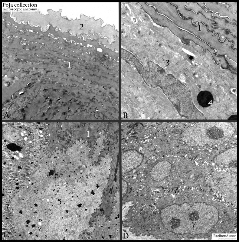
Title: Epithelial cell types in esophagus (gerbil, rat)
Description: Electron micrographs. (A, B, C) Gerbil. (D) Rat.
The esophagus of the gerbil and rat, in contrast to human, is to a certain extent keratinized, due to the type of food they eat.
(1) Stratum granulosum and (2) a “cleared” cell of the stratum lucidum. The dark aggregates represent the keratohyalin granules.
Note that these cell layers miss already the cell nuclei.
The layer below, shown in (B), still contains cells with nuclei, tonofilaments (3) and non-aggregated keratohyalin granules or melanin granules (4).
Similar layer, shown in (C), shows both small and round melanin grains, aggregated keratohyalin granules (6) and tonofilaments or keratin filaments (5). The basal cells show spikes or lateral interdigitations (7) between each other with many desmosomal connections (arrows,↘), to enforce resistance to the hard shell food, like nuts.
Keywords/Mesh: esophagus, squamous epithelium, keratinization, gerbil, melanin, electron microscopy, POJA collection