
1.1 POJA-L618. Eosinophilic granulocyte (peripheral blood, human)
|
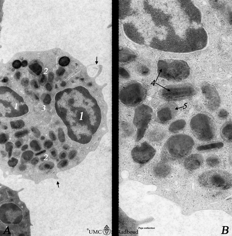
1.1 POJA-L618
Title: Eosinophilic granulocyte (peripheral blood, human) Description: Electron microscopy. (A): Overview of the cell. Note two nuclear lobes (1) due to the section. There are numerous specific eosinophilic granules (2) and a Golgi area (3). Few thin filopodia (↓, arrows) are present. (B): Detail: some vesicles and many specific eosinophilic granules of varying sizes. These granules contain a central electron-dense angular or round crystalloid core (4) embedded in a finely granular matrix (5). The crystalloid consists of an alkaline protein called major basic protein (MBP), the matrix contains eosinophilic cationic protein (ECP), lysosomal enzymes such as eosinophil peroxidase for the production of hypochlorous or hypobromous acid, lactoferrin and hydrolases. |
Background: Although this cell can internalise antigen-antibody complexes, their primary function is host defence against parasite infections. Eosinophils are normally present in peripheral tissues, especially in mucosal linings of the respiratory, gastrointestinal, and genitourinary tracts, and their numbers can increase by recruitment in the setting of inflammation. IL-5, released by Th2 cells enhances the ability of eosinophils to release granule contents on cross-linking of FcE-receptors. The granule proteins are toxic to parasites but may also injure normal tissue. Activated eosinophils, like mast cells and basophils, produce and release lipid mediators including PAF, prostaglandins and leukotrienes (LTC4, LTD4, LTE4). Also a variety of cytokines are produced by the eosinophils (IL-3, IL-5, IL-8, GM-CSF, IL-10 etc).
Keywords/Mesh: blood, bone marrow, eosinophilic granulocyte, specific granule, lysosome, phagocytosis, histology, electron microscopy, POJA collection