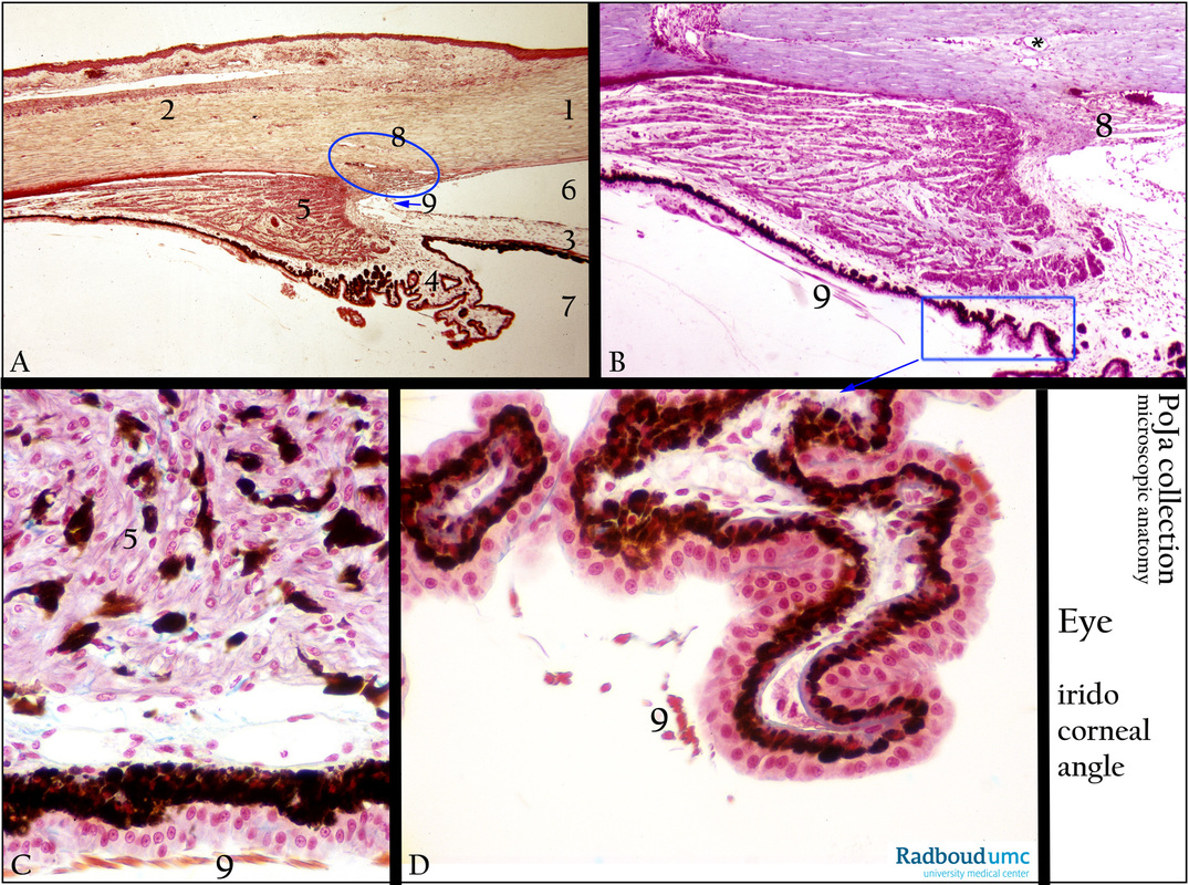
12.1.3 POJA-L2556+2557+2560+2559
Title: Ciliary body of the eye
Description:
(A, B): Survey ciliary body, stain Haematoxylin-Delafield, stain Azan respectively, human. (C, D): Ciliary body, stain Azan, monkey.
(1) Cornea.
(2) Sclera.
(3) Iris. Note that the iris (3) has a posterior double layer of epithelium of which the lower, outer layer is heavily pigmented.
(4) Ciliary body with ciliary processes. The pigmented layer of the two-layered epithelium in the ciliary body area, lining the ciliary processes however is located at the inner side facing the ciliary stroma (detail in C and D).
(5) Ciliary muscle.
(6) Anterior eye chamber.
(7) posterior eye chamber.
(8) (blue oval) Sinus venosus sclerae/limbus corneae.
(9) (blue arrow) Angulus iridocornealis. In the iridicorneal area arises a corneoscleral meshwork of trabeculae (8) which are a
loosely arranged sponge-like connective tissue, through which the ocular fluid flows towards the canal of Schlemm, detailed in (B, *).
The intertrabecular space is lined with flattened endothelium and is called the space of Fontana (B, 8), a space through which the
anterior chamber of the eye communicates with the canal of Schlemm.
A detail (B, blue rectangle) of the epithelium of the ciliary processes is shown enlarged in (D). (B, D, C, 9) represents the zonular fibres.
Contraction of the ciliary muscle results in reduction of the length of ciliary zonular fibres (= circular suspensory ligaments of the lens).
Note that in the monkey’s eye the ciliary smooth muscle contains many melanocytes (C).
Background: In human the ciliary muscle consists of interwoving muscle fibres with some regional variations in its fibre orientation.
The inner circular muscle fibres are parasympathetic innervated while the meridional-radial fibres (located closest to the sclera)
are sympathetic innervated.
Keywords/Mesh: eye, ciliary body, ciliary muscle, iridocorneal angle, Schlemm canal, histology, POJA collection

