
12.1.4 POJA-L2583+3821+
4428+2582
Optic nerve of the eye
12.1.4 POJA-L2583+3821+4428+2582
Title: Optic nerve of the eye
Description:
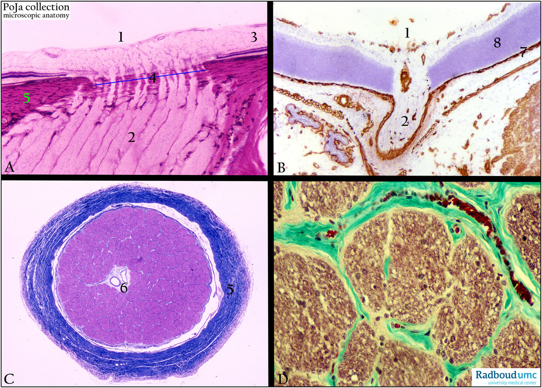
(A): Optic papilla, stain Haematoxylin-eosin, human. (1) Optic papilla with nonmyelinated axons (3) from the multipolar ganglion cells at the
inner side of the retina. The axons converge to the optic papilla to form the intraocular part of the optic nerve (2).
Subsequently it enters through the locally perforated sclera (5) and becomes myelinated and forms the orbital part of the
optic nerve. That perforated plate is called lamina cribrosa (4).
The optic disc includes the optic papilla (the protrusion formed by axons entering the optic nerve) and the lamina cribrosa of the sclera through which the axons pass.
(B): Optic nerve, immunoperoxidase staining with DAB and anti-collagen IV antibodies, 1day postnatal rat.
The basement membrane collagen IV is already observed only around blood vessels and capillaries at the inner side of the retina
and in the lamina basalis between the pigmented layer and the choriocapilaris area (7).
Note that in this area (8) no differentiation into inner and outer nuclear layers is yet observed.
In the postnatal rat close to the blank sclera bundles of striated ocular muscle fibres (right side) as well excretory glands (left side) are surrounded by collagen IV-positive basement membranes.
(C): Cross-section of optic nerve, stain Azan, human. In the cross-section distinctly blood vessels (6) are noticeable (arteria and vena centralis retinae). (5) Meninges, including dura mater, arachnoidea and pia mater.
(D): Cross-section of optic nerve, stain trichrome Goldner, human. It illustrates that the optic nerve is packed in myelinated cables surrounded with connective tissue sheaths (green).
Keywords/Mesh: eye, optic nerve, optic papilla, optic disc, lamina cribrosa, collagen IV, histology, POJA collection
Title: Optic nerve of the eye
Description:
(A): Optic papilla, stain Haematoxylin-eosin, human. (1) Optic papilla with nonmyelinated axons (3) from the multipolar ganglion cells at the
inner side of the retina. The axons converge to the optic papilla to form the intraocular part of the optic nerve (2).
Subsequently it enters through the locally perforated sclera (5) and becomes myelinated and forms the orbital part of the
optic nerve. That perforated plate is called lamina cribrosa (4).
The optic disc includes the optic papilla (the protrusion formed by axons entering the optic nerve) and the lamina cribrosa of the sclera through which the axons pass.
(B): Optic nerve, immunoperoxidase staining with DAB and anti-collagen IV antibodies, 1day postnatal rat.
The basement membrane collagen IV is already observed only around blood vessels and capillaries at the inner side of the retina
and in the lamina basalis between the pigmented layer and the choriocapilaris area (7).
Note that in this area (8) no differentiation into inner and outer nuclear layers is yet observed.
In the postnatal rat close to the blank sclera bundles of striated ocular muscle fibres (right side) as well excretory glands (left side) are surrounded by collagen IV-positive basement membranes.
(C): Cross-section of optic nerve, stain Azan, human. In the cross-section distinctly blood vessels (6) are noticeable (arteria and vena centralis retinae). (5) Meninges, including dura mater, arachnoidea and pia mater.
(D): Cross-section of optic nerve, stain trichrome Goldner, human. It illustrates that the optic nerve is packed in myelinated cables surrounded with connective tissue sheaths (green).
Keywords/Mesh: eye, optic nerve, optic papilla, optic disc, lamina cribrosa, collagen IV, histology, POJA collection