
14.2 POJA-L6120 Electron micrograph of a mitochondrion in cardiocytes (monkey)
14.2 POJA-L6120 Structure of mitochondrion in cardiocytes
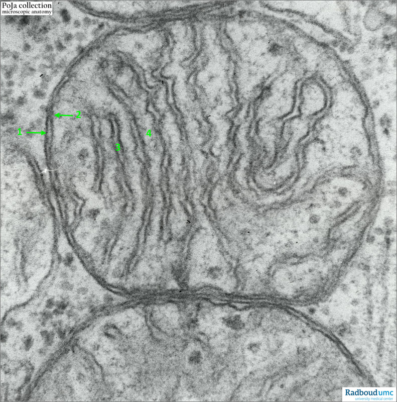
14.2 POJA-L6120 Electron micrograph of a mitochondrion in cardiocytes (monkey)
itle: Electron micrograph of a mitochondrion in cardiocyte (monkey)
Description:
The mitochondrion is composed of the outer mitochondrial membrane (OMM), the intermembrane space (between 1 and 2), the inner mitochondrial membrane (IMM) which is folded into projections called cristae, and the matrix. In cardiocytes of many mammals mitochondria exhibit more cristae that are better developed than those of mitochondria in skeletal or smooth muscle cells.
(1): Outer membrane
(2): Inner membrane
(3): Crista
(4): Matrix, with dispersed dense matrix granules containing a.o. calcium and magnesium
Mitochondria are double-membraned organelles which play important roles in cellular homeostasis, including producing energy by oxidative phosphorylation, maintaining calcium homeostasis, and regulating the signalling that leads to programmed cell death.
However, under stresses from environmental insults, mitochondrial shape, membrane potential and metabolism will be disturbed, and mitochondrial DNA, reactive oxidative species (ROS) and cytochrome c may be released into the cytoplasm. Moreover, mitochondrial proteins may even misfold under more severe conditions, causing a plethora of diseases such as cardiovascular disease (CVD), neurological diseases, aging and so on.
See also:
Keywords/Mesh: locomotor system, heart, ventricle, myocardiocyte, cardiocyte, myofilament, mitochondrion, electron microscopy, POJA collection
Description:
The mitochondrion is composed of the outer mitochondrial membrane (OMM), the intermembrane space (between 1 and 2), the inner mitochondrial membrane (IMM) which is folded into projections called cristae, and the matrix. In cardiocytes of many mammals mitochondria exhibit more cristae that are better developed than those of mitochondria in skeletal or smooth muscle cells.
(1): Outer membrane
(2): Inner membrane
(3): Crista
(4): Matrix, with dispersed dense matrix granules containing a.o. calcium and magnesium
Mitochondria are double-membraned organelles which play important roles in cellular homeostasis, including producing energy by oxidative phosphorylation, maintaining calcium homeostasis, and regulating the signalling that leads to programmed cell death.
However, under stresses from environmental insults, mitochondrial shape, membrane potential and metabolism will be disturbed, and mitochondrial DNA, reactive oxidative species (ROS) and cytochrome c may be released into the cytoplasm. Moreover, mitochondrial proteins may even misfold under more severe conditions, causing a plethora of diseases such as cardiovascular disease (CVD), neurological diseases, aging and so on.
See also:
- Review article: Mitochondrial Quality Control in Cardiomyocytes: A Critical Role in the Progression of Cardiovascular Diseases. Front. Physiol., 27 March 2020 | https://doi.org/10.3389/fphys.2020.00252
- 13.1 POJA-L4528+4529+4715+4530+4532+4533+4540
- 14.2 POJA-L6100+6104+6105 Cardiac muscle fibres with intercalating discs
- 14.2 POJA- POJA-L6099+14.2 POJA-L6099+6101+61066101+6106 Foetal and adult cardiocytes
- 13.1 POJA-L4510+4512 Lipofuscin in cardiocytes (ventricle, senium human
- 13.1 POJA-L4511+4513+4505+4514+4515 Lipofuscin and endocrine granules in myocardiocytes
- 13.1 POJA-L4517+4518+4519+4520+4521+4522 Myocardiocytes in foetal heart
- 13.1 POJA-L4526+4715+4527 Myocardiocytes
- 13.1 POJA-L4528+4529+4715+4530+4532+4533+4540 Electron micrographs of myocardiocytes
- 13.1 POJA-L4534+4536+4535+4539+4538 Purkinje fibres in the heart
- 13.1 POJA-L4735C+4729B+4503+4537+4501+4502 Macroscopy, microscopy and scheme of the heart
- 13.1 POJA-L4504+4531+4717+4716 Myocardiocytes
- 13.1 POJA-L4506+4509+4507+4508 Foetal and adult cardiocytes with lipofuscin (ventricle)
Keywords/Mesh: locomotor system, heart, ventricle, myocardiocyte, cardiocyte, myofilament, mitochondrion, electron microscopy, POJA collection