7.1 POJA-L1934+1935+1936+1938
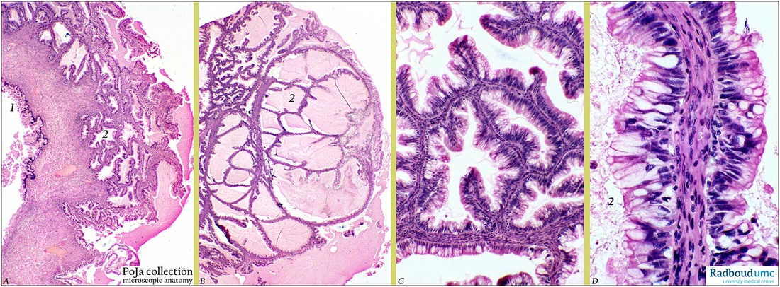
Title: Mucinous cystadenocarcinoma of ovary (human, adult)
Description: (A, B, C, D) Stain: Hematoxylin-eosin.
(A): Low magnification of well-differentiated mucinous adenocarcinoma with a papillary growth pattern. At (1) cystic formation.
(B): Another part of the tumor consists of glandular acini filled with mucoid material (2).
(C): Higher magnification of complex branching mucous tubules reveals lining of columnar epithelial cells.
(D): The mucus-secreting cells resemble those of the endocervix with tall columnar mucus-containing cell lining. Cellular atypia and nuclear stratification are present. Stroma (3) and mucoid secretion in lumen (2).
Background: Histogenetically it is assumed that a majority of epithelial tumors of the ovary is derived from the ovarian serosa. This lining is the equivalent of the embryonic coelomic epithelium in adulthood. Embryologically the coelomic epithelium give rise to the müllerian epithelium (ducts of Müller) and its derivates are the fallopian tube lining (tubal pathway), endometrial lining (endometrial pathway) and endocervical glands lining (endocervical pathway). The ovarian surface epithelium contains undifferentiated cells that still are latent to differentiate along similar embryonic pathways. In this way a tumor derived from these cells will differentiate in similar way. It appears that mucinous tumors are differentiated along the endocervical pathway. 10% of all ovarian cancers belong to the group of mucinous cystadenocarcinomas, and 20% of them are found bilaterally. Generally they appear as multiloculated tumors filled with a glycoprotein rich gelatinous fluid. In appearance they are similar to colonic neoplasms, but can be distinguished by their content of keratin 7 in contrast to normal colon and colon cancers. Histological the neoplasm presents an acinar or glandular pattern as well as solid sheets of cells with intracellular mucus. The solid growth shows more cellular atypia and stratification. Loss of glandular structure and areas of hemorrhage and necrosis are common. Pools of mucus might be formed in ovarian stroma due to seeping of mucus out from any mucinous tumor resulting in the so-called pseudomyxoma ovarii.
Keywords/Mesh: female reproductive organs, ovary, female genitalia, , ovarian neoplasms, mucinous cystadenocarcinoma, histology, POJA collection

