
14.3 POJA-L6133+6134 α-Smooth actin staining of the internal anal sphincter (pig)
14.3 POJA-L6133+6134 α-Smooth actin immunoperoxidase staining of the internal anal sphincter
14.3 POJA-L6133+6134 α-Smooth actin staining of the internal anal sphincter (pig)
Title: α-Smooth actin staining of the internal anal sphincter (pig)
Description:
The musculus sphincter ani encloses the anal canal and is divided in an internal part and an external part. The internal is a continuation of the circular muscle layer of the rectum and is thickened as the non-striated sphincter ani internus (involuntary muscle). The external part or the sphincter ani externus (voluntary muscle) is composed of skeletal muscles and encompasses the internal part. It is subdivided in three parts from bottom to the top: a deep part (pars profunda) close to the internal part, followed by a superficial part located beneath the profunda and a subcutaneous part under the skin at the anal side. In these areas smooth and striated muscles are found close together.
The use of a smooth actin antibody (a-SMA, alpha-SM-actin or ACTA2) is an easy and quick marker to detect the presence of intact, damaged or deformed muscular part of the internal anal sphincter.
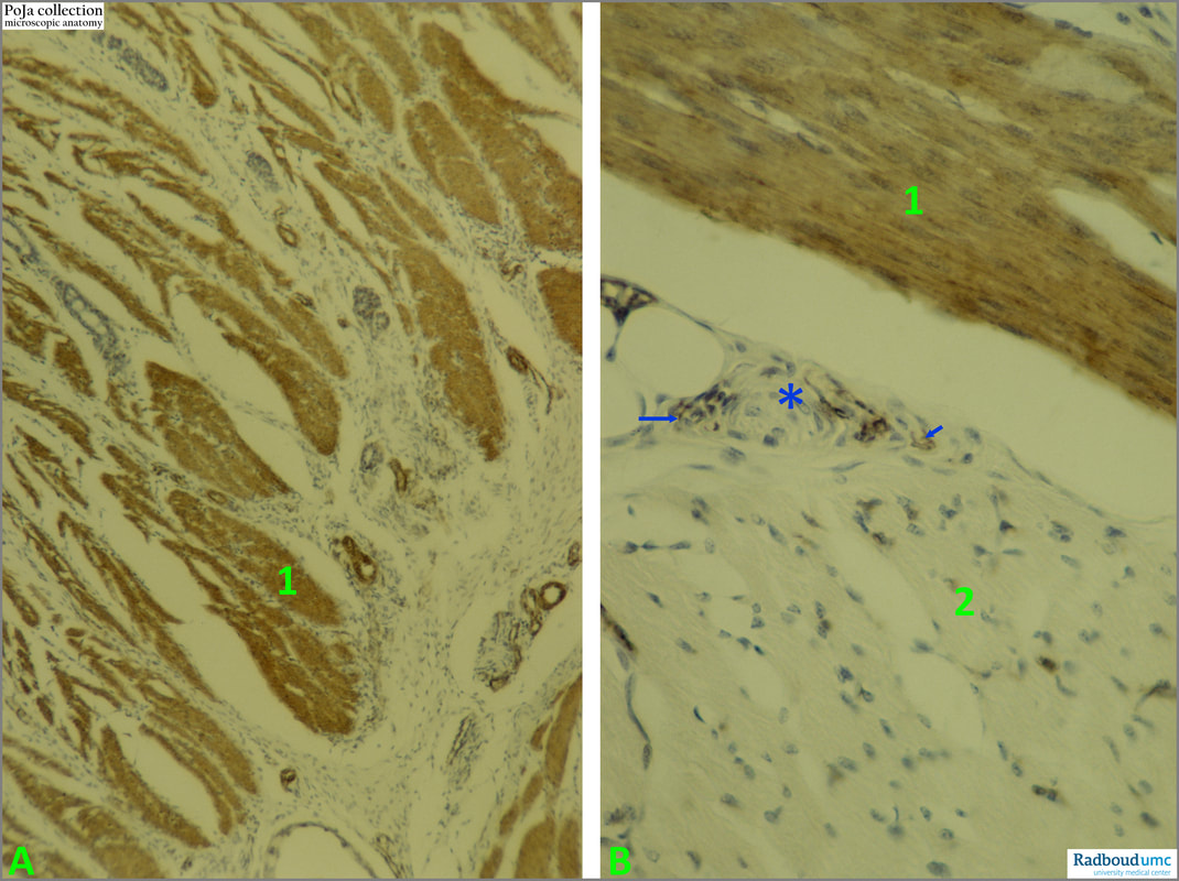
(A): Survey, (B): Detail.
(A): Note that the internal anal sphincter (1) is a smooth muscle type which is stained positively. Small circular structures are blood vessels with positive smooth muscles in their walls (media). Glandular tubules (anal glands) are negative with exception of the positive myoepithelial cells that contain smooth actin filaments.
(B): The differentiation is facilitated between the positive internal sphincter muscle cells (1) and the external sphincter (2) that is composed of a striated muscle type completely negative for α-smooth actin. The actins are a family of globular proteins that form microfilaments and the α-SMA is one of 6 different actin isoforms in smooth muscle. The isoform of actin ACTA1 (or Alpha-actin-1) only expressed in striated muscle (skeletal and cardiac tissue) differs 8 amino acids from the α-SMA and does not react with smooth actin antibodies.
At (arrows) capillaries with positive pericytes and (*) indicates a sensory receptor (lamellar body) present in perimysium of skeletal muscle. Still a fine striation of the myofibres is detectable.
Keywords/Mesh: locomotor system, skeletal muscle, striated muscle, smooth muscle, anal sphincter, actin, ACTA2, a-smooth actin, a-SMA, alpha-SM-actin, histology, POJA-collection
Description:
The musculus sphincter ani encloses the anal canal and is divided in an internal part and an external part. The internal is a continuation of the circular muscle layer of the rectum and is thickened as the non-striated sphincter ani internus (involuntary muscle). The external part or the sphincter ani externus (voluntary muscle) is composed of skeletal muscles and encompasses the internal part. It is subdivided in three parts from bottom to the top: a deep part (pars profunda) close to the internal part, followed by a superficial part located beneath the profunda and a subcutaneous part under the skin at the anal side. In these areas smooth and striated muscles are found close together.
The use of a smooth actin antibody (a-SMA, alpha-SM-actin or ACTA2) is an easy and quick marker to detect the presence of intact, damaged or deformed muscular part of the internal anal sphincter.
(A): Survey, (B): Detail.
(A): Note that the internal anal sphincter (1) is a smooth muscle type which is stained positively. Small circular structures are blood vessels with positive smooth muscles in their walls (media). Glandular tubules (anal glands) are negative with exception of the positive myoepithelial cells that contain smooth actin filaments.
(B): The differentiation is facilitated between the positive internal sphincter muscle cells (1) and the external sphincter (2) that is composed of a striated muscle type completely negative for α-smooth actin. The actins are a family of globular proteins that form microfilaments and the α-SMA is one of 6 different actin isoforms in smooth muscle. The isoform of actin ACTA1 (or Alpha-actin-1) only expressed in striated muscle (skeletal and cardiac tissue) differs 8 amino acids from the α-SMA and does not react with smooth actin antibodies.
At (arrows) capillaries with positive pericytes and (*) indicates a sensory receptor (lamellar body) present in perimysium of skeletal muscle. Still a fine striation of the myofibres is detectable.
Keywords/Mesh: locomotor system, skeletal muscle, striated muscle, smooth muscle, anal sphincter, actin, ACTA2, a-smooth actin, a-SMA, alpha-SM-actin, histology, POJA-collection