
6.3 POJA-L4243+2735+
2740+2738.
Seminal vesicles
6.3 POJA-L4243+2735+2740+2738
Title: Seminal vesicles
Description:
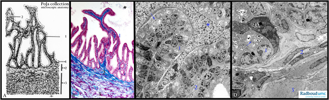
(A) Scheme. (B) Stain Azan, human. (C, D) Electron microscopy, gerbil.
(A): Shows that the structure of the seminal vesicle (or glandula vesiculosa) in fact is a coiled tube with a folded mucosa creating invaginations or a labyrinth-like structure. (1) A two-layered epithelium (1), with secretory activity, detailed in (C, D) with arrows
pointing to vesicles. The lumen (*) between the folds in (B, C) is filled with secreted liquid, pigment and cellular remnants.
Close to the base of the epithelium is located the proper lamina with stroma cells (A, B, D, 2) followed by a well-developed muscle
layer of helical winded smooth muscles (A, B, 3) detailed in (D, 3).
Keywords/Mesh: seminal vesicle, seminal gland, glandula vesiculosa, histology, electron microscopy, POJA collection
Title: Seminal vesicles
Description:
(A) Scheme. (B) Stain Azan, human. (C, D) Electron microscopy, gerbil.
(A): Shows that the structure of the seminal vesicle (or glandula vesiculosa) in fact is a coiled tube with a folded mucosa creating invaginations or a labyrinth-like structure. (1) A two-layered epithelium (1), with secretory activity, detailed in (C, D) with arrows
pointing to vesicles. The lumen (*) between the folds in (B, C) is filled with secreted liquid, pigment and cellular remnants.
Close to the base of the epithelium is located the proper lamina with stroma cells (A, B, D, 2) followed by a well-developed muscle
layer of helical winded smooth muscles (A, B, 3) detailed in (D, 3).
Keywords/Mesh: seminal vesicle, seminal gland, glandula vesiculosa, histology, electron microscopy, POJA collection