
4.1.1 POJA-L4210. Esophagus and transition into cardia-area (human)
4.1.1 POJA-L4210
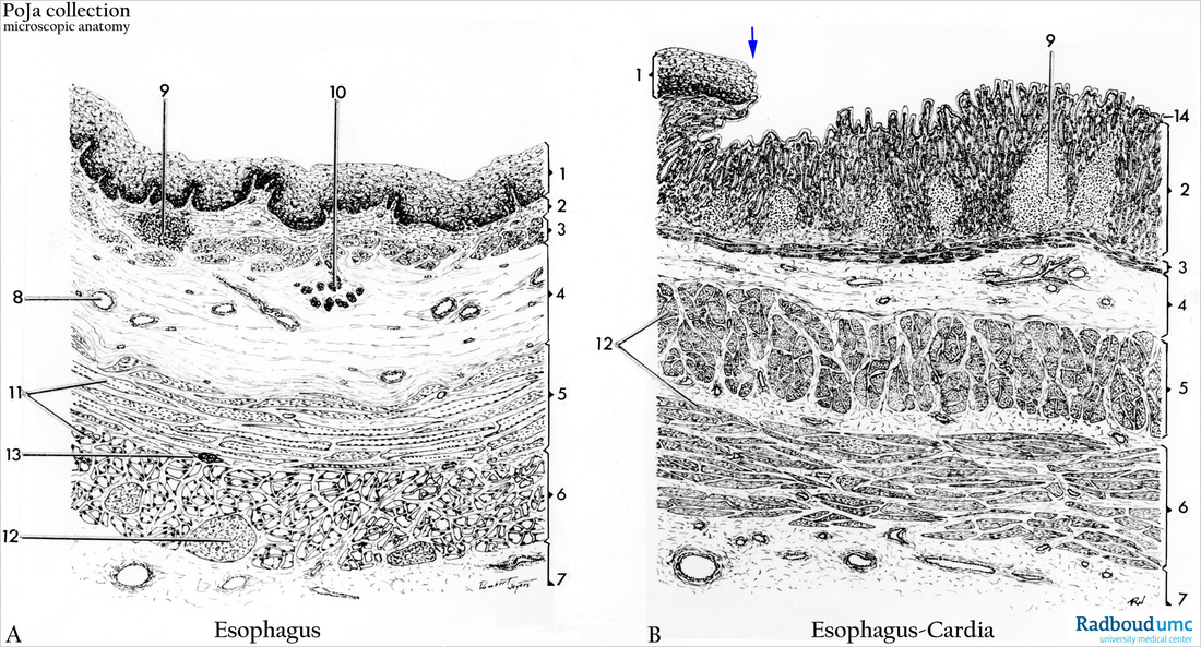
Title: Esophagus and transition into cardia-area (human)
Description: Light microscopy scheme. (A) Transvers section esophagus. (B) Longitudinal section transition zone esophagus- cardia.
(1, A, B) Lamina epithelialis mucosae, multilayered non-keratinizing squamous epithelium.
(2, A, B) Lamina propria, with glands in (B);
(3, A, B) Lamina muscularis mucosae;
(4, A, B) Tela submucosa. Layers 1-4 together are called tunica mucosa.
(5, A, B) Inner circular muscle layer or stratum circulare;
(6, A, B) Outer longitudinal muscle layer or stratum longitudinale. Both layers together are called tunica muscularis.
(7, A, B) Tunica adventitia.
(8, A ) Venous plexus in the tela submucosa.
(9 A, B) Folliculus lymphaticus solitaries.
(10, A ) Mucinous glandulae oesophageae.
(11, A ) Striated muscles.
(12, A, B) Smooth muscles.
(13, A ) Plexus myentericus (Auerbach).
(14, B ) Single layered columnar epithelium. At (arrow,↘) in (B) the transition between squamous epithelium and single columnar epithelium is marked.
Background: The main function of the esophagus is transport of food to the stomach. The epithelium, being continuous with the oral cavity lining, consists of non-keratinizing squamous epithelium supported with numerous connective tissue papillae and being mechanically resistant to the food passage. The muscular layer of the mucosa is of the skeletal striated type near the pharynx but gradually downwards turns into smooth muscle fibers. Two types of glands are present in the esophagus: (1) the glandulae propriae or glandulae oesophageae in the submucosa layer throughout the entire esophagus. They are tubulo-alveolar glands and produce lubricating mucus. (2) The cardiac glands situated in the lamina propria just below the lining epithelium in the lower 1/3 part of the esophagus near the transition zone to the stomach. They have elaborately branched tubules in irregular shapes and produce mucus. At the end of the pars cardiaca, at the entrance of the stomach, the squamous esophageal epithelium abruptly passes into a single layered columnar epithelium of the stomach. There the mucosa forms invaginations (foveolae and crypts) and the character of the slightly curling cardiac glands turn into stomach glands with different types of cells. Whereas the lamina muscularis mucosae regulates the mobility of the mucosa, the tunica muscularis, surrounding the whole esophagus, takes care for the movement of the food towards the stomach. The double muscle layer consists of an inner circular layer and an outer longitudinal layer, in between them provided with several nerve plexus. A loose connective tissue layer surrounds the esophagus and facilitates a solid but flexible position of the esophagus and conducts blood vessels and nerve fibers.
Keywords/Mesh: esophagus, cardia, squamous epithelium, columnar epithelium, stomach, histology, POJA collection