
4.1.1 POJA-L-4018+4019+La0169. Plica, villi and crypts in the jejunum (human)
4.1.1 POJA-L4018+4019+La0169
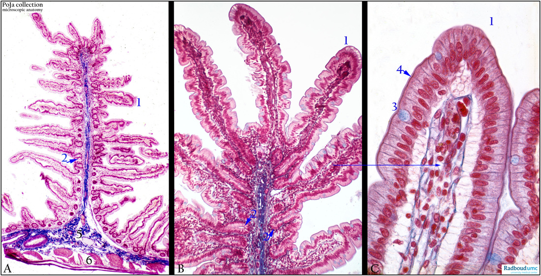
Title: Plica, villi and crypts in the jejunum (human)
Description: Stain: Azan.
(A): Kerckring’s plica or fold formed by mucosa and submucosa layer (5, blue). The tunica muscularis (6) is not involved in the plica formation. The fold is further amplified by the presence of villi (1) and crypts (2).
(B, C): Details of a plica with villi. The core of a villus is supported by lamina propria connective tissue that embeds capillaries and small lymph vessels as shown in (C). Note that bluish goblet cells (3) are present between the columnar epithelial cell linings. The latter are provided with a brush border (4) as further surface amplification. The major function of the surface epithelial cells is the intake of digested food elements, while the goblet cells are in charge of producing mucus.
Keywords/Mesh: jejunum, plicae, villi, crypts, histology, POJA collection