
12.1.4 POJA-L2566+4421
Retina, periphery and parafoveal area
12.1.4 POJA-L2566+4421
Title: Retina, periphery and parafoveal area
Description:
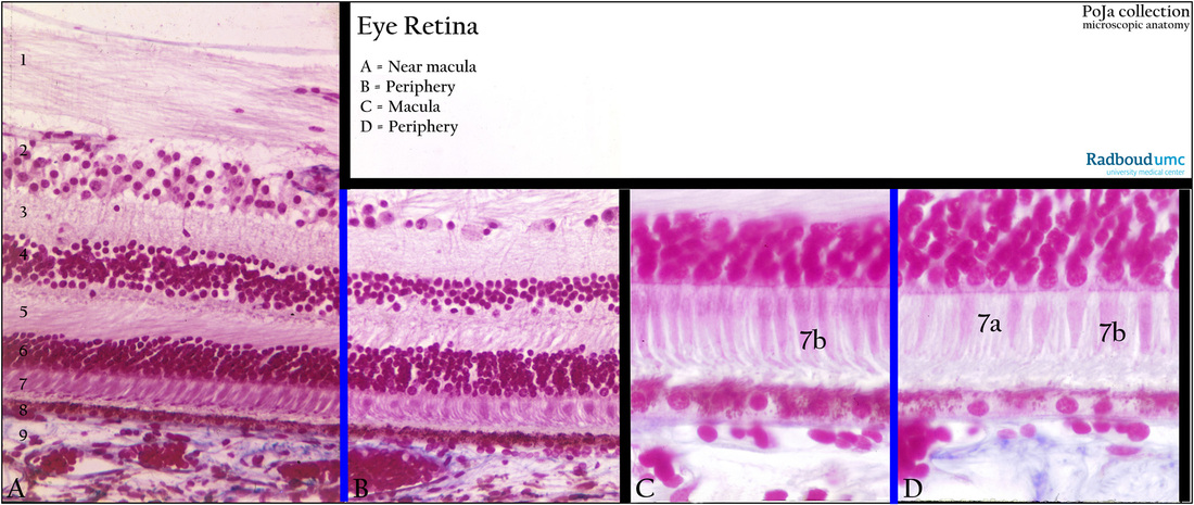
(A, C): Macula site (parafoveal area) of the retina, stain Azan, human.
(B, D): Peripheral area of the retina, stain Azan, human.
The sensory retina contains cones (A, 7 and D, 7b) and rods (D, 7a) with exception of the optic papilla (optic disc) and the centre of the foveola.
Background: At the posterior pole of the eye a yellow spot about 1.5 mm diameter is called the macula lutea due to local accumulation
of exogenous xanthophyll (carotenoid) pigments.
It is located about 4 mm temporal to the optic papilla and the pigments absorb short wavelight light.
This macula lutea has an annular pigmented ridge caused by an accumulation of nerve cells bipolar ganglion cells and nerve fibres.
The central part is a small depression i.e. fovea centralis the thinnest part of the retina.
The inner centre of the fovea is the foveola characterised by the presence of only Müller cells.
(A) versus (B), Comparable Identical layers:
(1) Nerve fibres layer, thick in A.
(2) Ganglion cell layer, thicker in A.
(3) Inner plexiform layer.
(4) Inner nuclear layer.
(5) Outer plexiform layer (oblique fibres).
(6) Outer nuclear layer.
(7) Inner and outer segments of cones and rods.
(8) Retinal pigmented epithelium (RPE).
(9) Choriocapillaris.
(C) versus (D):
Photoreceptor layer: Parafoveal area versus peripheral area.
In contrast to the rods (D, 7a) responsible for dim light vision and movement detection, the cones (D, 7b) are involved in color
distinction and acute vision. Especially in the fovea (centre of the macula) exclusively long slender cones (C, 7b) are present as photoreceptor cells and thus the fovea is for high quality vision.
These cones synapse with bipolar cells. Both, however, are oriented at an angle around the margins of the foveola so that the inner
nuclear and ganglion layers are displaced somewhat toward the outer limiting membrane. In this way the outer nuclear layer and
all other layers don’t obstruct the light pathway. Only Müller cells between the cones are present.
Keywords/Mesh: eye, retina, macula lutea, fovea centralis, rod, cone, Müller cell, histology, POJA collection
Title: Retina, periphery and parafoveal area
Description:
(A, C): Macula site (parafoveal area) of the retina, stain Azan, human.
(B, D): Peripheral area of the retina, stain Azan, human.
The sensory retina contains cones (A, 7 and D, 7b) and rods (D, 7a) with exception of the optic papilla (optic disc) and the centre of the foveola.
Background: At the posterior pole of the eye a yellow spot about 1.5 mm diameter is called the macula lutea due to local accumulation
of exogenous xanthophyll (carotenoid) pigments.
It is located about 4 mm temporal to the optic papilla and the pigments absorb short wavelight light.
This macula lutea has an annular pigmented ridge caused by an accumulation of nerve cells bipolar ganglion cells and nerve fibres.
The central part is a small depression i.e. fovea centralis the thinnest part of the retina.
The inner centre of the fovea is the foveola characterised by the presence of only Müller cells.
(A) versus (B), Comparable Identical layers:
(1) Nerve fibres layer, thick in A.
(2) Ganglion cell layer, thicker in A.
(3) Inner plexiform layer.
(4) Inner nuclear layer.
(5) Outer plexiform layer (oblique fibres).
(6) Outer nuclear layer.
(7) Inner and outer segments of cones and rods.
(8) Retinal pigmented epithelium (RPE).
(9) Choriocapillaris.
(C) versus (D):
Photoreceptor layer: Parafoveal area versus peripheral area.
In contrast to the rods (D, 7a) responsible for dim light vision and movement detection, the cones (D, 7b) are involved in color
distinction and acute vision. Especially in the fovea (centre of the macula) exclusively long slender cones (C, 7b) are present as photoreceptor cells and thus the fovea is for high quality vision.
These cones synapse with bipolar cells. Both, however, are oriented at an angle around the margins of the foveola so that the inner
nuclear and ganglion layers are displaced somewhat toward the outer limiting membrane. In this way the outer nuclear layer and
all other layers don’t obstruct the light pathway. Only Müller cells between the cones are present.
Keywords/Mesh: eye, retina, macula lutea, fovea centralis, rod, cone, Müller cell, histology, POJA collection