
7.2 POJA-L1808B+1818+1816+1822. Cervical intraepithelial neoplasm (CIN 3) (human, adult)
7.2 POJA-L1808B+1818+1816+1822
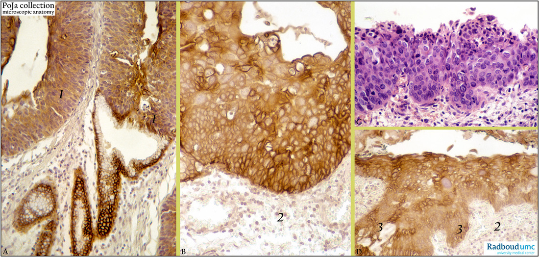
Title: Cervical intraepithelial neoplasm (CIN 3) (human, adult)
Description: Stain: (C) Hematoxylin-eosin; (A, B, D) CK 7 antikeratin 7 antibody (OVTL 12-30) immunoperoxidase staining with diaminobenzidin reaction (DAB) and hematoxylin counterstaining.
(A): CK 7-positive immature squamous epithelium (1), extending abnormal epithelium into a mucous gland.
(B): The full thickness of immature squamous epithelium shows heterogeneity in strong CK 7 expression. Stroma (2) is negative.
(C): Mitoses through full thickness of epithelium; Note high nucleus/cytoplasmic (N/C) ratio of the cells and loss of polarity.
(D): CK 7-positive squamous cell hyperplasia ; (3) with extensions down into the CK 7-negative stroma (2).
(Partly by courtesy of G.P. Vooijs MD PhD, former Head of the Department of Pathology, Radboud university medical center, Nijmegen, The Netherlands).
Clinical background: The proliferation of keratine-7 positive reserve cells, near the squamocolumnar junction, leads to a multilayer. With increasing maturation this layer shows squamous differentiation resulting in squamous metaplasia and note that these cells still preserve their CK 7- reactivity. Immunohistological analysis of these reserve cells have indeed proven the presence of cytokeratins of both columnar epithelial cells (low molecular weight keratin) as well as of non-keratinised squamous epithelial cells (high molecular weight keratin). Apparently reserve cells possess the potential towards a dual differentiation. The CIS cells preserve during squamous metaplasia their cytokeratin profile of the basal reserve cells.
Keywords/Mesh: female reproductive organs, cervix, CIS, cervical intraepithelial neoplasia (CIN), carcinoma in situ (CIS), uterine cervical neoplasms, cytokeratin 7, histology, POJA collection.