
10.3 POJA-L2053+2233+
2237+2250+2232+2231.
Vascularization in dermis
10.3 POJA-L2053+2233+2237+2250+2232+2231
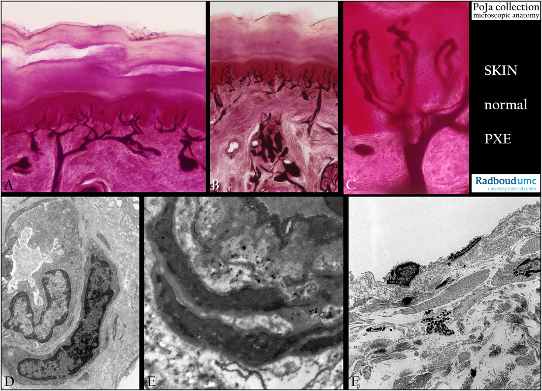
Title: Vascularization in dermis
Description:
Indian ink perfusion-eosin to illustrate the delicate vascularization in the papillary layer of the fingertip (A) and nail bed (B, C), human.
Electron micrographs of (D) Normal blood capillary in a muscle biopsy, human.
(E) Abnormal electron-dense calcium deposits subendothelially and deeper in an arteriole. Skin of belly, pseudoxanthoma elasticum. This disorde is a genetic disease (autosomal recessive mutation) causing fragmentation and mineralization (calcification) of elastic fibers in skin and eyes, blood vessels.
(F) Normal lymph capillary in breast skin, human. Note distinct bundles of collagen fibers and most likely
a mast cell in close vicinity.
Keywords/Mesh: skin, vascularization, blood capillary, lymph capillary, pseudoxanthoma elasticum, histology, electron microscopy, POJA collection