
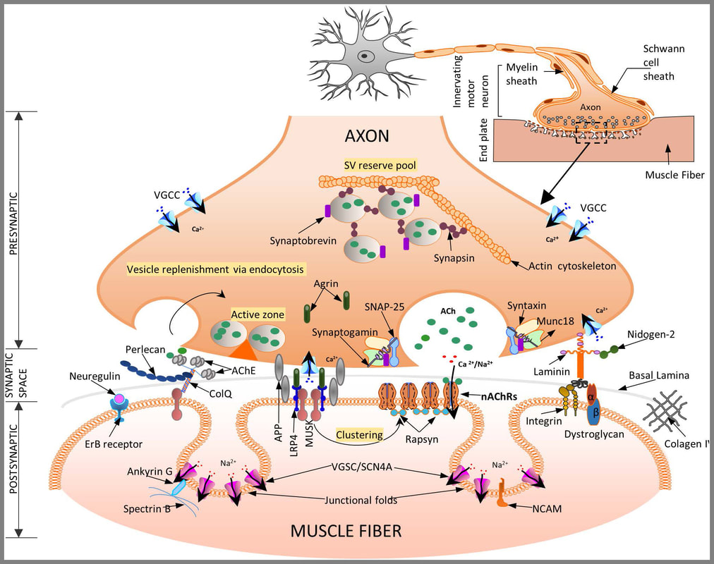
14.1 POJA-L6294-B Synapse scheme of a neuromuscular junction (NMJ)
14.1 POJA-L6294-B Synapse scheme of a neuromuscular junction
14.1 POJA-L6294-B Synapse scheme of a neuromuscular junction (NMJ)
(Skeletal muscle: A review of molecular structure and function, in health and diseaseKavitha Mukund and Shankar Subramaniam, First published: 13 August 2019, https://doi.org/10.1002/wsbm.1462 ) © 2019 The Authors. WIREs Systems Biology and Medicine published by Wiley Periodicals, Inc. This is an open access article under the terms of the Creative Commons Attribution License, which permits use, distribution and reproduction in any medium, provided the original work is properly cited.
Title: Synapse scheme of a neuromuscular junction (NMJ)
Description:
The structure of the neuromuscular junction consists of
Nerve Terminal: The motor neuron terminal forms a complex of 100-200 branching nerve endings, called nerve terminals.
The nerve terminal membrane has areas of membrane thickening called active zones. Local active zones have a family of SNAP proteins (syntaxins and synaptosome-associated protein 25) and rows of voltage-gated calcium (Ca) channels. The nerve terminal also has K-channels on its membrane. The cytoplasm contains mitochondria, endoplasmic reticulum, and synaptic vesicles (SVs). Each SV stores around 5000-10000 molecules of acetylcholine (ACh), the neurotransmitter at Neuro Muscular Junction (NMJ). The SVs are concentrated around the active zone. The membrane of SVs has synaptogmin and synaptobrevin proteins, being essential for fusion and docking of SVs at active zones. Upon an action potential Ca channels open to cause influx, which induces concentration of SVs at active zones and exocytosis of the ACh from the synaptic vesicles into the synaptic cleft.
The synaptic cleft measures ∼50 nm and contains nicotinic ACh receptors on the motor endplate site. The synaptic cleft contains acetylcholinesterase enzyme responsible for the catabolism of released ACh so that its effect on the post-synaptic receptors is not prolonged.
The motor endplate forms the postsynaptic part of NMJ. It is the thickened portion of the sarcolemma that is folded into junctional folds. They have ACh receptors concentrated at the top of the folds. They function as Ach-gated ion channels. Binding of ACh to these receptors opens the channels allowing the influx of Na-ions from the extracellular fluid into the muscle membrane. This creates endplate potential and generates and transmits AP to the muscle membrane.
Summarizing: A schematic representation of a neuromuscular junction (NMJ) and its main molecular actors—three specific regions define the NMJ:
(a): The presynaptic motor nerve terminal where vesicles fuse with the terminal membrane to release acetylcholine (ACh) into the synaptic
cleft. Calcium influx through the voltage-gated Ca channels (VGCC) trigger vesicle fusion and release from the active zones.
(b): The synaptic space contains the basal lamina (BL, extra cellular matrix layer), and shows the presence of AChEsterase-COLQ (essential for
the inactivation of ACh). Acetylcholine esterase is anchored to the BL via COLQ and perlecan, being essential for stabilization of BL. MuSK enables AChR clustering via rapsyn.
(c): Postsynaptic organization of the skeletal muscle membrane include several folds with receptors for the diffusing ACh (AChRs) at the crest and voltage-gated sodium channels (VGSC) in the troughs of the folds necessary for efficient neuromuscular transmission. The agrin-Lrp4-MuSK complex, present on the trough of the postsynaptic membrane is essential for the formation of the NMJ. Agrin, a NMJ heparan sulphate (HS) proteoglycan (PG) is critical for organization of the Ach receptors and NMJ.
The entire structure is finally attached to the actin cytoskeleton (not shown here for simplicity).
For a detailed review of the contribution of all proteins involved in the signal transduction mechanism we refer to the article: Skeletal; muscle: A review of molecular structure and function, in health and disease, by K. Mukund and S. Subramaniam (Wire’s Syst.Biol.Med. 2020;12: e 1462) (https://doi.org/10.1002/wsbm.1462).
See also:
Keywords/Mesh: locomotor system, skeletal muscle, striated muscle, synapse, motor endplate, myoneural junction, neuromuscular junction, scheme, POJA collection
Title: Synapse scheme of a neuromuscular junction (NMJ)
Description:
The structure of the neuromuscular junction consists of
- The presynaptic part (nerve terminal),
- The postsynaptic part (motor endplate),
- The synaptic cleft, between nerve terminal and motor endplate.
Nerve Terminal: The motor neuron terminal forms a complex of 100-200 branching nerve endings, called nerve terminals.
The nerve terminal membrane has areas of membrane thickening called active zones. Local active zones have a family of SNAP proteins (syntaxins and synaptosome-associated protein 25) and rows of voltage-gated calcium (Ca) channels. The nerve terminal also has K-channels on its membrane. The cytoplasm contains mitochondria, endoplasmic reticulum, and synaptic vesicles (SVs). Each SV stores around 5000-10000 molecules of acetylcholine (ACh), the neurotransmitter at Neuro Muscular Junction (NMJ). The SVs are concentrated around the active zone. The membrane of SVs has synaptogmin and synaptobrevin proteins, being essential for fusion and docking of SVs at active zones. Upon an action potential Ca channels open to cause influx, which induces concentration of SVs at active zones and exocytosis of the ACh from the synaptic vesicles into the synaptic cleft.
The synaptic cleft measures ∼50 nm and contains nicotinic ACh receptors on the motor endplate site. The synaptic cleft contains acetylcholinesterase enzyme responsible for the catabolism of released ACh so that its effect on the post-synaptic receptors is not prolonged.
The motor endplate forms the postsynaptic part of NMJ. It is the thickened portion of the sarcolemma that is folded into junctional folds. They have ACh receptors concentrated at the top of the folds. They function as Ach-gated ion channels. Binding of ACh to these receptors opens the channels allowing the influx of Na-ions from the extracellular fluid into the muscle membrane. This creates endplate potential and generates and transmits AP to the muscle membrane.
Summarizing: A schematic representation of a neuromuscular junction (NMJ) and its main molecular actors—three specific regions define the NMJ:
(a): The presynaptic motor nerve terminal where vesicles fuse with the terminal membrane to release acetylcholine (ACh) into the synaptic
cleft. Calcium influx through the voltage-gated Ca channels (VGCC) trigger vesicle fusion and release from the active zones.
(b): The synaptic space contains the basal lamina (BL, extra cellular matrix layer), and shows the presence of AChEsterase-COLQ (essential for
the inactivation of ACh). Acetylcholine esterase is anchored to the BL via COLQ and perlecan, being essential for stabilization of BL. MuSK enables AChR clustering via rapsyn.
(c): Postsynaptic organization of the skeletal muscle membrane include several folds with receptors for the diffusing ACh (AChRs) at the crest and voltage-gated sodium channels (VGSC) in the troughs of the folds necessary for efficient neuromuscular transmission. The agrin-Lrp4-MuSK complex, present on the trough of the postsynaptic membrane is essential for the formation of the NMJ. Agrin, a NMJ heparan sulphate (HS) proteoglycan (PG) is critical for organization of the Ach receptors and NMJ.
The entire structure is finally attached to the actin cytoskeleton (not shown here for simplicity).
For a detailed review of the contribution of all proteins involved in the signal transduction mechanism we refer to the article: Skeletal; muscle: A review of molecular structure and function, in health and disease, by K. Mukund and S. Subramaniam (Wire’s Syst.Biol.Med. 2020;12: e 1462) (https://doi.org/10.1002/wsbm.1462).
See also:
- 14.1 POJA-L6304B Electron micrograph of a motor endplate on skeletal muscle fibre
- 11.2.2 POJA-L3278+3297+ 4434+4435+4436+4437 Neuromuscular junction (peripheral ending of effector neuron
Keywords/Mesh: locomotor system, skeletal muscle, striated muscle, synapse, motor endplate, myoneural junction, neuromuscular junction, scheme, POJA collection