6.1 POJA-L2657+3345
Title: Embryonic and juvenile testes
Description:
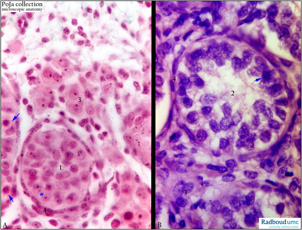
Stain hematoxylin-eosin, human. (A) Embryonic testis. (B) Juvenile testis (1.5 years).
In the seminiferous tubule (A, 1) the Sertoli cells (*) with a clearly depicted nucleolus are visible (small arrows).
In both sections (A, 1 and B, 2), however, intermediate cells of the spermatogenesis are yet not present,
except for some spermatogonial cells (arrows↘↘). (3) Interstitial Leydig cells.
(4) Boundary lining (lamina propria limitans) of the tubular wall.
Keywords/Mesh: testis, fetus, postnatal, Sertoli cell, Leydig cell, spermatogonium, histology, POJA collection

