
10.3 POJA-L2037+2242+
2080+2087+2243+2089.
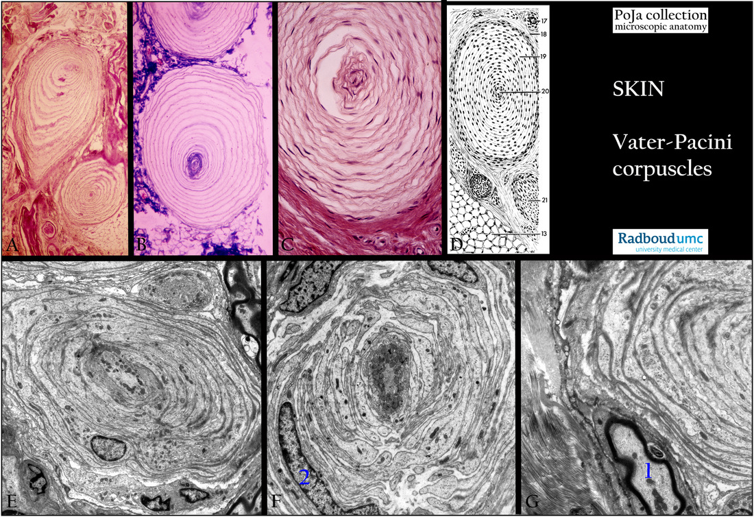
Vater-Pacini corpuscles I in the skin
10.3 POJA-L2037+2242+2080+2087+2243+2089
Title: Vater-Pacini corpuscles I in the skin
Description:
Vater-Pacini corpuscles (pacinian corpuscles).
(A): Foot sole, stain hematoxylin-azophloxine, human.
(B): Foot sole, stain Azan, human.
(C): Fingertip, stain hematoxylin-azophloxine, human.
(D): Light microscopy scheme, human:
(17) Small artery.
(18) Connective tissue sheath (capsule).
(19) Perineural lamellae.
(20) Thickened nerve ending.
(21) Nerve bundle.
(13) Fatty tissue.
(E - G): Snout skin, electron microscopy, pig.
The Vater-Pacini corpuscles are large lamellated corpuscles found in the subcutaneous tissue of the palms of the hand,
soles of the foot and other places as tendons and pancreas.
The sheaths of the nerve endings are characteristically arranged in lamellae as in onion shells (perineural lamellae).
The afferent axon nerve fiber is located in the center and is extremely rich in mitochondria (E, F).
They function as pressure and vibration receptors. (G,1) Myelinated axon. (F, 2) Modified Schwann cell.
The pacinian corpuscle functions as a transducer which responds to mechanical vibrations, pressure and tensions.
The lamellae and the interlamellar space that is filled with liquid probably amplify any distortion or movement and
thus improve the signal.
Keywords/Mesh: skin, Vater-Pacini corpuscle, pacininan corpuscle, sensoric receptor, histology, electron microscopy, POJA collection