
12.2.4.2 POJA-L3423+3402+
2635+2634+2636
Crista ampullaris in the inner ear
12.2.4.2 POJA-L3423+3402+2635+2634+2636
Title: Crista ampullaris in the inner ear
Description:
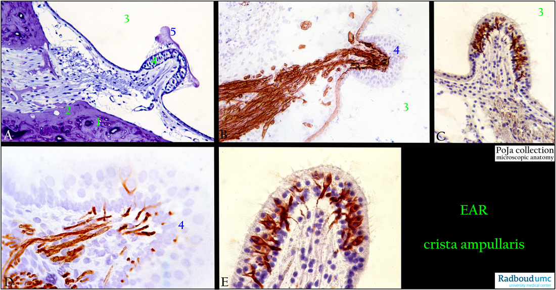
(A): Ampulla with crista ampullaris, stain toluidine blue, semi-thin plastic section, rat. In the adult rat the bony labyrinth is characterised
by cross-sections of the Haversian systems or osteons formed by circular arranged osteocytes (1).
The peripheral part is composed of the inner circumferential bone lamellae (2). [For bone see also 16.1.3 POJA-L7112+7074+7117 Lamellar bone (or secondary bone)]
In a dilated part of a semicircular canal or the ampulla (3) myelinated nerve fibres (branch of the vestibular nerve) penetrate the vascularised loose connective tissue (perilymphatic tissue) up to the bottom of the ridge (crista ampullaris) where supporting and
sensory cells (neuroepithelium, 4) are located.
Remnants of the jelly-like cupula (5) are shown due to histological preparation procedures. At the base of the crista the supporting cells are continuous with the flattened cells in this area (planum semilunatum).
(B): Crista ampullaris, immunoperoxidase staining with AEC and antibodies against collagen IV, 4 days postnatal rat.
Note the nerve fibre bundle (utriculoampullar nerve) marked by the basal lamina containing collagen IV as well as several capillaries
and the lining of the membranous ampulla.
(C, D, E): Crista ampullaris, immunoperoxidase staining with AEC and antibodies against neurofilament, 4 days and 10days postnatal rats. The fine nerve fibre branches penetrate the neuroepithelium and form a basket of connections with and around the sensory hair cells.
Keywords/Mesh: inner ear, vestibular organ, semicircular canal, crista ampullaris, collagen IV, neurofilament, planum semilunatum. histology, POJA collection
Title: Crista ampullaris in the inner ear
Description:
(A): Ampulla with crista ampullaris, stain toluidine blue, semi-thin plastic section, rat. In the adult rat the bony labyrinth is characterised
by cross-sections of the Haversian systems or osteons formed by circular arranged osteocytes (1).
The peripheral part is composed of the inner circumferential bone lamellae (2). [For bone see also 16.1.3 POJA-L7112+7074+7117 Lamellar bone (or secondary bone)]
In a dilated part of a semicircular canal or the ampulla (3) myelinated nerve fibres (branch of the vestibular nerve) penetrate the vascularised loose connective tissue (perilymphatic tissue) up to the bottom of the ridge (crista ampullaris) where supporting and
sensory cells (neuroepithelium, 4) are located.
Remnants of the jelly-like cupula (5) are shown due to histological preparation procedures. At the base of the crista the supporting cells are continuous with the flattened cells in this area (planum semilunatum).
(B): Crista ampullaris, immunoperoxidase staining with AEC and antibodies against collagen IV, 4 days postnatal rat.
Note the nerve fibre bundle (utriculoampullar nerve) marked by the basal lamina containing collagen IV as well as several capillaries
and the lining of the membranous ampulla.
(C, D, E): Crista ampullaris, immunoperoxidase staining with AEC and antibodies against neurofilament, 4 days and 10days postnatal rats. The fine nerve fibre branches penetrate the neuroepithelium and form a basket of connections with and around the sensory hair cells.
Keywords/Mesh: inner ear, vestibular organ, semicircular canal, crista ampullaris, collagen IV, neurofilament, planum semilunatum. histology, POJA collection