
11.4 POJA-L3108+4451+
3112+2975+3117+2968+2972+2976.
Cell types of the cerebellum
11.4 POJA-L3108+4451+3112+2975+3117+2968+2972+2976
Title: Cell types of the cerebellum
Description:
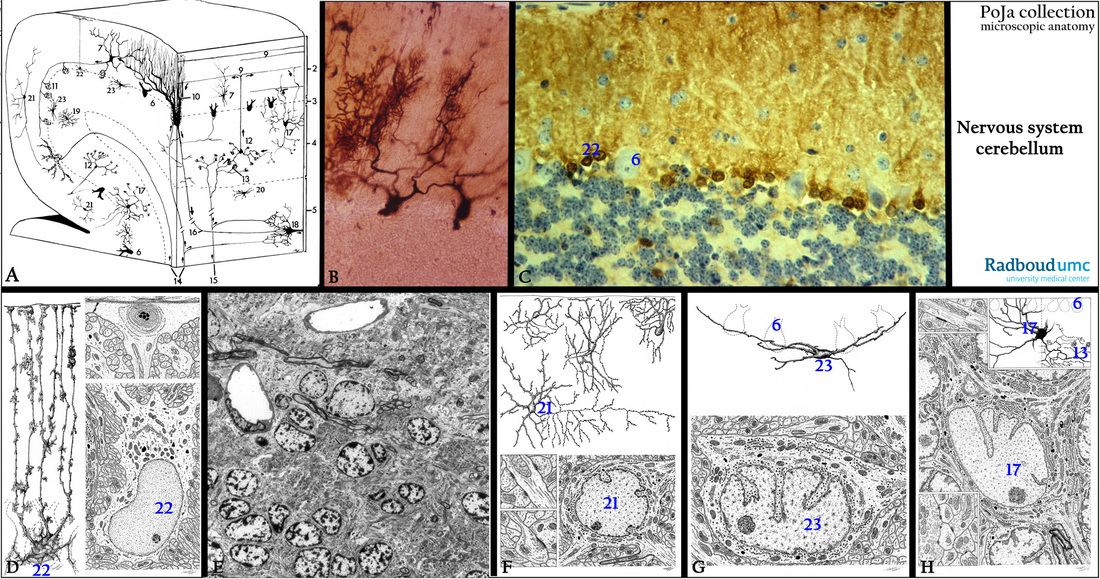
(A): Scheme cerebellum, human (zoom in).
Referred to Section 11.1.
(1) Pia mater.
(2) Molecular layer.
(3) Ganglion layer with Purkinje cells.
(4) Granular (or granule) layer.
(5) Medulla rich in myelinated fibers (white matter).
(6) Purkinje cells.
(7) basket cell (inhibitory).
(8) absent in this scheme
(9) Parallel fibers (axons of granule cells).
(10) Dendrites of Purkinje cells in contact with climbing fibers.
(11) Fibers of basket cell around Purkinje cell.
(12) Granule cells (excitatory neuron).
(13) Glomerulus (cerebellar island), contact place between mossy fibers and granule cells.
(14) Climbing fibers (input) ending on the proximal dendrites of Purkinje cells.
(15) Mossy fibers (input) ending in glomerulus.
(16) Axon of Purkinje cell (output from the cerebellar cortex).
(17) Golgi cell (inhibitory interneuron).
(18) Neuron in deep cerebellar nucleus.
(19) Astrocyte in granular layer. The protoplasmic asterocytes are present in the gray matter and their terminal feet end upon capillaries at one side and thus involved in the blood-brain-barrier (BBB) by formation of perivascular endfeet (membrana gliae limitans perivascularis). At the other side they end up on neurons.
(20) Astrocyte in medulla. These fibrous asterocytes are found in the white matter and just like the protoplasmic asterocytes their processes also form several perivascular endfeet or subpial endfeet.
(21) Outer stellate cell. The cerebellar stellate neuron is a multipolar interneuron with variable length of their processes and found in the molecular cortex.
(22) Bergmann glial cell. This cell is a specialized semi-radial glia cell that closely interacts with neuronal elements in the molecular layer of the cerebellum. With its long processes it contributes to the formation of the glial limiting membrane.
(23) Lugaro cell. An inhibitory interneuron which appears GABA-positive in the neuritic and somatic profiles. These cells seem to exert a feed-back inhibitory control on Purkinje cells.
(B): Golgi stain, human. In a partly sagittal/transversal section Purkinje cells with their dendritic arborizations
in the molecular layer.
(C): Immunoperoxidase staining with DAB and antibodies against S100 protein, mouse. Generally strong
positive reaction in the soma and processes of astroglial cells and Bergmann glial cells (22).
A more diffuse positive reactivity is found in the molecular layer due to the intricate network of glial processes
as well as the long processes of the Bergmann cells till the surface. Negative reaction in the Purkinje cell
somata (6), cerebellar glomeruli and granule interneurons in the granular layer.
(D): Electron microscopy scheme of the Bergman astrocytes (22), human. Bergmann glial cells are also called
‘specialized’ astrocytes closely interacting with neurons in the molecular layer. They have small somata with
up to 6 very long processes extend from the Purkinje cell layer to the pial surface and contribute to the formation
of the glia limitans (membrana gliae limitans superficialis). The elaborated processes of these glia cells form
close contacts with synapses of parallel fibers on the Purkinje cell dendritic branches. As electrically nonexcitable
cells they function in the same way as the protoplasmic astrocytes being responsible for glutamate uptake
and extracellular K+ homeostasis. Uptake of K+ is regulated by the glia cell cytosolic Ca2+ and provide a control mechanism of the membrane potentials and activity of regional Purkinje cells.
(E): Electron micrograph of the granular layer with the neuropil comprising of glomeruli, astroglial cells and myelinated axons, perfusion fixation, rabbit. Myelinated nerve fibers are recognizable as densely stained fibers.
(F): Electron microscopy scheme of stellate cells (21), human. Cerebellar stellate neurons are multipolar interneurons in the molecular layer and two types are distinguished. One with long axons running in a sagittal
plane, the other with short axons branching within the region dendritic projections.
(G): Electron microscopy scheme of Lugaro cell (23), human. This cell is an inhibitory interneuron located in
the granular layer closely below the Purkinje cell layer. It has a fusiform soma with thick horizontally oriented dendrites and axon processes in the molecular layer. The Lugaro cell appears GABA-positive in axons and soma. These axons form multiple synapses with the somata of basket and stellate cells, and possibly also with Golgi cell dendrites in the molecular layer. It is assumed that Lugaro cells exert a feed-back inhibitory control on Purkinje cells (6).
(H): Electron microscopy scheme of Golgi cell (17), human. The cerebellar Golgi neuron (cerebellar golgi cell) is
an interneuron located in the granule layer. Its dendrites extend into the molecular layer where they contact
with parallel fibers.
The axons ramifiy in the granule layer and form a complex cerebellar glomerulus (13) with granule cell dendrites
and mossy fiber terminals.
Keywords/Mesh: nervous tissue, cerebellum, pia mater, interneuron, Purkinje cell, Bergman glia cell, Lugaro cell, Golgi cell, stellate cell, astrocyte, S100, histology, electron microscopy, POJA collection
Title: Cell types of the cerebellum
Description:
(A): Scheme cerebellum, human (zoom in).
Referred to Section 11.1.
(1) Pia mater.
(2) Molecular layer.
(3) Ganglion layer with Purkinje cells.
(4) Granular (or granule) layer.
(5) Medulla rich in myelinated fibers (white matter).
(6) Purkinje cells.
(7) basket cell (inhibitory).
(8) absent in this scheme
(9) Parallel fibers (axons of granule cells).
(10) Dendrites of Purkinje cells in contact with climbing fibers.
(11) Fibers of basket cell around Purkinje cell.
(12) Granule cells (excitatory neuron).
(13) Glomerulus (cerebellar island), contact place between mossy fibers and granule cells.
(14) Climbing fibers (input) ending on the proximal dendrites of Purkinje cells.
(15) Mossy fibers (input) ending in glomerulus.
(16) Axon of Purkinje cell (output from the cerebellar cortex).
(17) Golgi cell (inhibitory interneuron).
(18) Neuron in deep cerebellar nucleus.
(19) Astrocyte in granular layer. The protoplasmic asterocytes are present in the gray matter and their terminal feet end upon capillaries at one side and thus involved in the blood-brain-barrier (BBB) by formation of perivascular endfeet (membrana gliae limitans perivascularis). At the other side they end up on neurons.
(20) Astrocyte in medulla. These fibrous asterocytes are found in the white matter and just like the protoplasmic asterocytes their processes also form several perivascular endfeet or subpial endfeet.
(21) Outer stellate cell. The cerebellar stellate neuron is a multipolar interneuron with variable length of their processes and found in the molecular cortex.
(22) Bergmann glial cell. This cell is a specialized semi-radial glia cell that closely interacts with neuronal elements in the molecular layer of the cerebellum. With its long processes it contributes to the formation of the glial limiting membrane.
(23) Lugaro cell. An inhibitory interneuron which appears GABA-positive in the neuritic and somatic profiles. These cells seem to exert a feed-back inhibitory control on Purkinje cells.
(B): Golgi stain, human. In a partly sagittal/transversal section Purkinje cells with their dendritic arborizations
in the molecular layer.
(C): Immunoperoxidase staining with DAB and antibodies against S100 protein, mouse. Generally strong
positive reaction in the soma and processes of astroglial cells and Bergmann glial cells (22).
A more diffuse positive reactivity is found in the molecular layer due to the intricate network of glial processes
as well as the long processes of the Bergmann cells till the surface. Negative reaction in the Purkinje cell
somata (6), cerebellar glomeruli and granule interneurons in the granular layer.
(D): Electron microscopy scheme of the Bergman astrocytes (22), human. Bergmann glial cells are also called
‘specialized’ astrocytes closely interacting with neurons in the molecular layer. They have small somata with
up to 6 very long processes extend from the Purkinje cell layer to the pial surface and contribute to the formation
of the glia limitans (membrana gliae limitans superficialis). The elaborated processes of these glia cells form
close contacts with synapses of parallel fibers on the Purkinje cell dendritic branches. As electrically nonexcitable
cells they function in the same way as the protoplasmic astrocytes being responsible for glutamate uptake
and extracellular K+ homeostasis. Uptake of K+ is regulated by the glia cell cytosolic Ca2+ and provide a control mechanism of the membrane potentials and activity of regional Purkinje cells.
(E): Electron micrograph of the granular layer with the neuropil comprising of glomeruli, astroglial cells and myelinated axons, perfusion fixation, rabbit. Myelinated nerve fibers are recognizable as densely stained fibers.
(F): Electron microscopy scheme of stellate cells (21), human. Cerebellar stellate neurons are multipolar interneurons in the molecular layer and two types are distinguished. One with long axons running in a sagittal
plane, the other with short axons branching within the region dendritic projections.
(G): Electron microscopy scheme of Lugaro cell (23), human. This cell is an inhibitory interneuron located in
the granular layer closely below the Purkinje cell layer. It has a fusiform soma with thick horizontally oriented dendrites and axon processes in the molecular layer. The Lugaro cell appears GABA-positive in axons and soma. These axons form multiple synapses with the somata of basket and stellate cells, and possibly also with Golgi cell dendrites in the molecular layer. It is assumed that Lugaro cells exert a feed-back inhibitory control on Purkinje cells (6).
(H): Electron microscopy scheme of Golgi cell (17), human. The cerebellar Golgi neuron (cerebellar golgi cell) is
an interneuron located in the granule layer. Its dendrites extend into the molecular layer where they contact
with parallel fibers.
The axons ramifiy in the granule layer and form a complex cerebellar glomerulus (13) with granule cell dendrites
and mossy fiber terminals.
Keywords/Mesh: nervous tissue, cerebellum, pia mater, interneuron, Purkinje cell, Bergman glia cell, Lugaro cell, Golgi cell, stellate cell, astrocyte, S100, histology, electron microscopy, POJA collection