7.3 POJA-L1379+1390
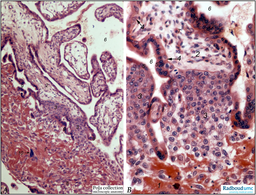
Title: Anchoring villi (human placenta, early midpregnancy)
Description: Stain: Hematoxylin –azophloxine. Survey (A) and detail (B).
The maternal side at the bottom shows reddish matrix-type fibrinoid accumulations (1) (Rohr’s fibrinoid layer) close to an anchoring villus (2) and the cytotrophoblastic cell columns (3). The anchoring villus is lined by cytotrophoblasts (4) which are covered at the outer side by multinuclear syncytiotrophoblasts (5). Intervillous space is marked by (6). (See also POJA-L1227).
(7) is a phagocytic Hofbauer cell,
(8) infiltrating extravillous cytotrophoblast cells between the fibrinoid deposits;
(9) points to the trophoblasts of the cytotrophoblast shell that covers part of the anchoring villus (3).
Arrow (10) indicates a multinuclear trophoblast cell that develops into a placental bed giant cell.
Keywords/Mesh: placenta, chorionic villi, Hofbauer cell, anchoring villus, cytotrophoblast, trophoblast, histology, POJA collection.

