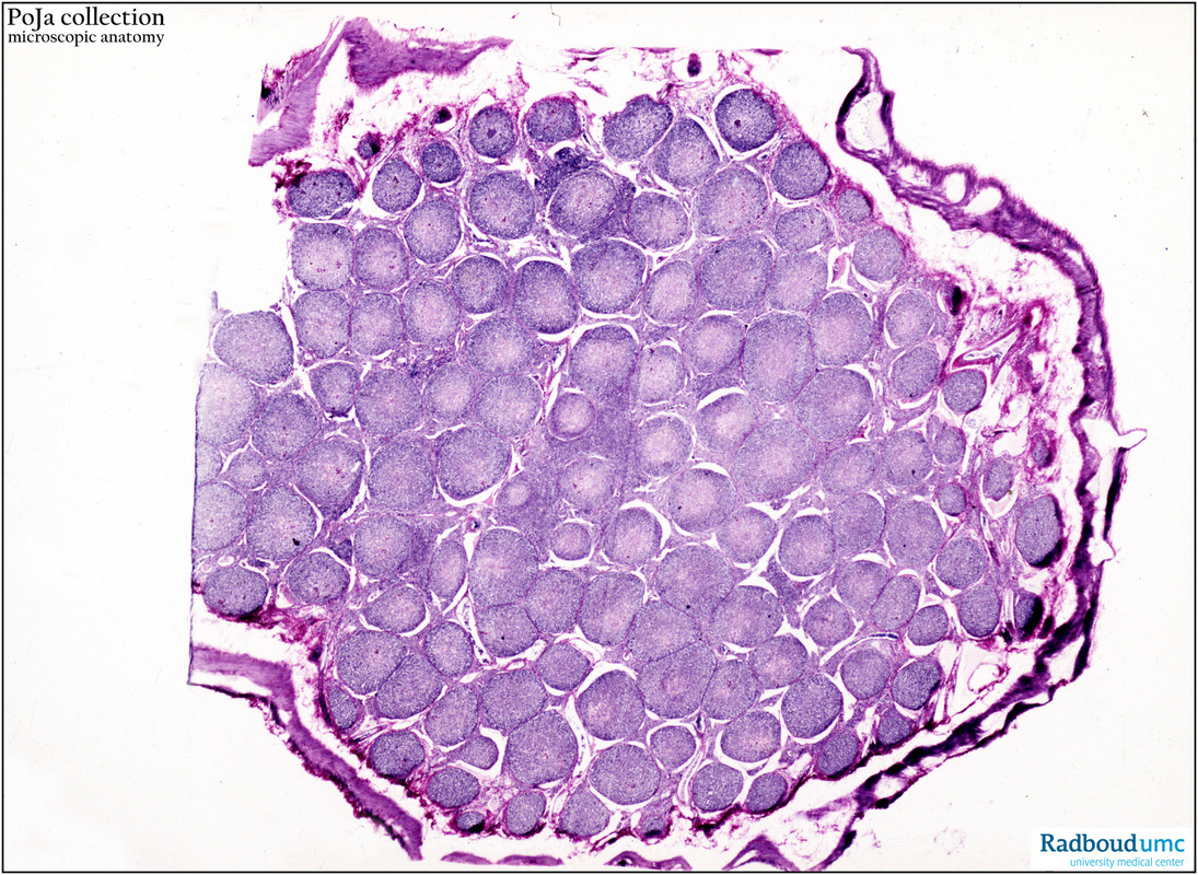
4.1.1 POJA-L4074
Title: Tangential section through Peyer’s patches in the ileum (dog)
Description: Stain: Hematoxylin-eosin.
The Peyer’s patches consist of aggregates of lymph follicles located in the submucosa and lamina propria of the ileum. They are macroscopically visible as white lucent knobs in or on the wall of the ileum. This tangential cut shows all the follicles centrally while rests of the wall of the digestive tract are left at the edge of the figure.
Keywords/Mesh: ileum, Peyer’s patches, histology, POJA collection

