
4.1.1 POJA-L3911+3918+3920+3921+3922. Keratin expression pattern in fetal and adult esophagus (human)
4.1.1 POJA-L-3911+3918+3920+3921+3922
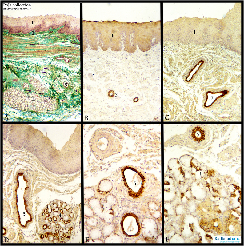
Title: Keratin expression pattern in fetal and adult esophagus (human)
Description: Stain: (A) Goldner (adult). (B-F) Anti-keratin antibody immunoperoxidase staining with diaminobenzidin reaction (DAB) and hematoxylin counterstaining.
(A): The cross section of the adult esophagus shows a multilayered non-keratinizing squamous epithelial covering (1) of the lumen, attached to a loose connective tissue layer (green), the so called lamina propria (2) and a small lamina muscularis mucosae (3) needed to adjust the movement of the epithelial layer around the lumen.
(4) Esophageal glands located in the submucosal layer ending in the corresponding duct (5). These glands in the upper one third of the esophagus are considered as continuations of the oral salivary glands. In this section the tunica muscularis and the adventitia layer are omitted.
(B, C): Stained with a poly-antikeratin serum reveals that both the epithelial linings plus the glandular ducts are visualized, glands however are not yet developed at the fetal stage here.
In (D-F) both the tubule acinar esophageal glands and the ducts are stained positively with anti keratin-7 monoclonal antibody (OVTL12/30). However, while all ductal cells are stained homogenously positive, the gland cells appear rather heterogeneously at this stage.
Note that keratin 7 is not present in the squamous lining epithelium.
Keywords/Mesh: esophagus, fetus, keratin, epithelium, histology, POJA collection