2.3 POJA-L1015
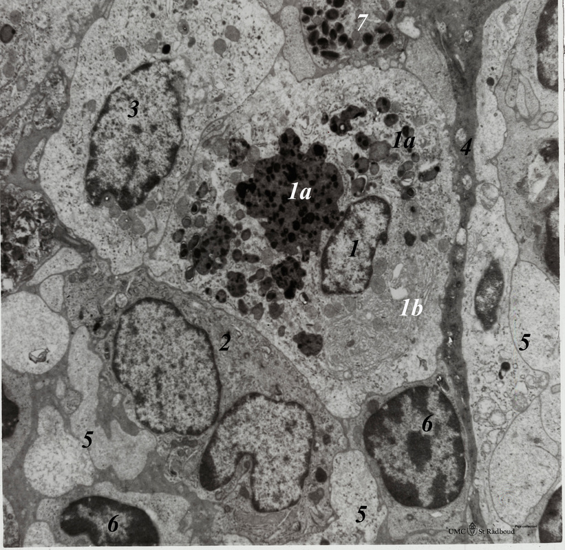
Title: Lymph node (rat)
Description: Electron microscopy.
Within the reticular framework of lymph nodes a variety of active reticular cells are present, some of them (1) have phagocytised foreign material (1a) and contain numerous lysosomal structures (1a) and large Golgi areas (1b)). Others are less active (2).
(3): Wandering interdigitating cell with its branches (5);
(4): Small electron-grey projection of a fibroblastic reticular cell;
(6): Lymphocytes;
(7): Eosinophilic granulocyte.
Keywords/Mesh: lymphatic tissue, lymph node, reticular tissue, reticular cell, interdigitating cell, phagocytosis, histology, electron microscopy, POJA collection

