
12.1.5 POJA-L2590+3819+
3563
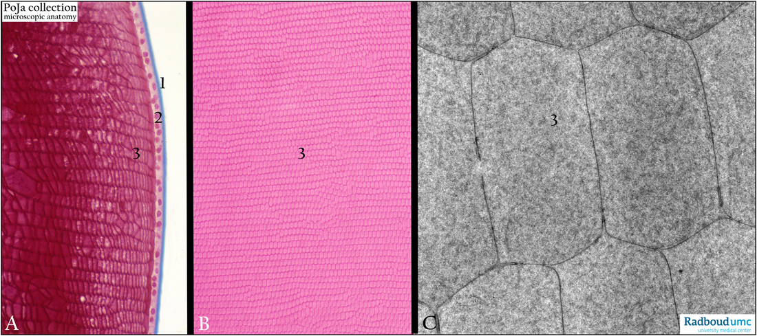
Lens fibres of the eye
12.1.5 POJA-L2590+3819+3563
Title: Lens fibres of the eye
Description:
(A): Lens, stain Azan, monkey. (1) Lens capsule consisting of collagen IV and glycosaminoglycans. (2) One layer of the anterior lens epithelium. (3) Note that the lens fibres (elongated lens cells) have lost their nuclei.
(B, C): Lens, stain Haematoxylin-eosin, pig (B). Electron micrograph, bovine (C).
Details of the hexagonal lens fibres (anterior cortex). Note ultrasturcturally the uniform aspect of the granular cytoplasm,
adjacent cells are separated by a thin intercellular space (about 15 nm width).
Background: Lens fibres filled with several types of proteins e.g. filensin (IF filament protein that contains attachment sites
for crystallins) and lens-specific proteins the α-, β-, γ-crystallins.
Both proteins maintain the conformation and transparency of the lens fibre cells.
Keywords/Mesh: eye, lens, lens fibre, histology, electron microscopy, POJA collection