13.1 POJA-L4528+4529+4715+4530+4532+4533+4540
Title: Electron micrographs of myocardiocytes
Description:
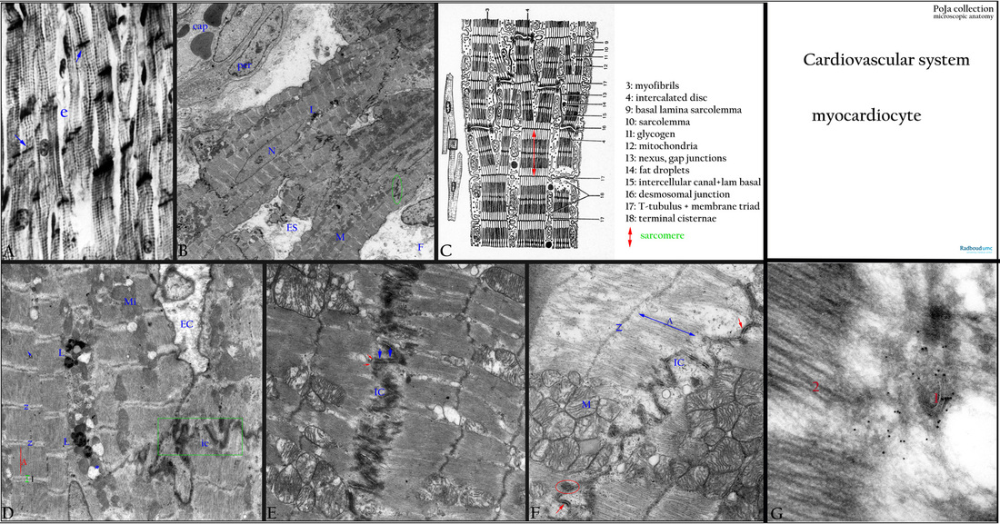
(A): Heart atrium, cardiocytes, black white, human. The striated cells show alternating dark A-bands and I-bands. The nucleus is located centrally in the cell. Numerous intercalated discs as anchoring sites of branching myocardiocytes (arrows). (e) Thin endomysium.
(B): Heart ventricle, cardiocytes, EM, rat. Survey of about six anastomosing myocardiocytes with left a capillary (cap) with erythrocytes and surrounded by pericyte (per). (N) Central nucleus of a cardiocyte with paranuclearly small lipofuscin granules (L) and mitochondria (M). One of several intercalated discs is encircled. (ES) extracellular space. (F) A damaged fibroblast. Transverse tubules of the T-system are present forming a small triad with terminal endings of the sarcoplasmic reticulum at the level of each Z-line [not visible in this survey; the reader is referred to Locomotor System in section 14.1 POJA-L6227-B Electron micrograph of the sarcomere structure in myofibres (human), POJA-L6248 Electron micrograph of the triad structure in striated muscle (human)].
(C): Scheme EM cardiocyte with sarcomere configuration of the myofilaments.
(D): Heart ventricle, cardiocytes, EM, rat. Detail of (B) with two cells linked together by an intercalated disc (IC), lipofuscin granules (L), normal mitochondria (Mi) and ballooning (*) mitochondria (in the process of degeneration). Z-lines (Z), A-bands (A), small I-bands (I) (contraction of myofilaments) and faintly M-bands (M) are present. (EC) extracellular space.
(E): Heart ventricle, cardiocytes, EM, human. Two cells linked together by an intercalated disc (IC), the disc is a transverse structure that occurs at the level of the Z-lines and represents an area where the cardiocytes and their branches meet end-to-end. The intercalated disc is differentiated into zones of desmosome (macula adhaerens) (thick blue arrows), intermediate junction (fascia adhaerens) and (thin red arrows) gap-junction (nexus).
(F): Heart ventricle, cardiocytes, EM, human. One damaged cell linked to another one by an intercalated disc (IC). Aggregated swollen mitochondria (M) (biopsy during operation). Z-lines (Z), double headed arrow: location of A-band (A). Macula adhaerens (red arrows) and fascia adhaerens (red oval).
(G): Heart ventricle, cardiocyte, immunoelectron microscopy, mouse. Ultra-cryosection shows black dots of gold-labeled desmin localised in the region of the macula adhaerens (1) as well as in the cytoskeletal structures near the thick myosin filaments (2).
Background: Laterally the junctions of the disc are not in register but arranged as steps of a ‘staircase’. In certain areas the surface of apposed cell membranes parallels the long axis of the cell. Here longer stretches of gap-junctions (or nexuses) will be present (vertical part of the steps in the ‘staircase’). As a specialised junctional complex the intercalated discs are involved in cell cohesion, in transmitting tension and contraction impulses form cell to cell. With ageing they become more complex and increase in amount.
Keywords/Mesh: cardiovascular system, heart, ventricle, myocardiocyte, cardiocyte, myofibril, myofilament, desmin, sarcomere, intercalated disc, sarcoplasmic reticulum, T-tubulus, triad, lipofuscin, electron microscopy, histology, POJA collection

