
5.4.1 POJA-L2544+2472+
2536+2535.
Podocytes in glomerulus (VII) of the kidney
5.4.1 POJA-L2544+2472+2536+2535
Title: Podocytes in glomerulus (VII) of the kidney
Description:
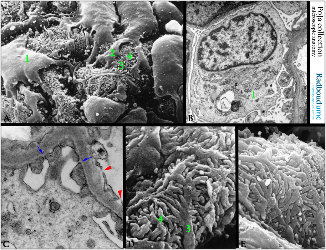
(A): Podocytes (1), scanning electron microscopy, human. Long primary processes (2), branch into secondary ones (3) and subsequently
into tertiary processes (4).
(B): Electron microscopy of a podocyte (1) with an elaborate Golgi area, a large lysosomal structure, human. Broad foot processes on the glomerular basal lamina (GBL), even in normal GBL, splitting is observed. Generally the GBL is composed of three layers a dense layer squeezed between inner and outer light layers: a lamina rara interna (LRI), lamina densa (LD) and a lamina rara externa (LRE).
(C): Detail of the pedicles and the ultrafiltration slit (or filtration slit membrane, blue arrows) between them.
The gap between them varies between 30 and 60 nm. The endothelial cell cytoplasm is fenestrated (red arrowheads).
(D, E): Scanning electron microscopy of the pedes or pedicles, the thick finger-like processes of the human podocyte e.g. secondary
processes (3) and tertiary processes (4).
Keywords/Mesh: urinary system, kidney, glomerulus, podocyte, filtration slit membrane, pedicle, histology, electron microscopy, POJA collection
Title: Podocytes in glomerulus (VII) of the kidney
Description:
(A): Podocytes (1), scanning electron microscopy, human. Long primary processes (2), branch into secondary ones (3) and subsequently
into tertiary processes (4).
(B): Electron microscopy of a podocyte (1) with an elaborate Golgi area, a large lysosomal structure, human. Broad foot processes on the glomerular basal lamina (GBL), even in normal GBL, splitting is observed. Generally the GBL is composed of three layers a dense layer squeezed between inner and outer light layers: a lamina rara interna (LRI), lamina densa (LD) and a lamina rara externa (LRE).
(C): Detail of the pedicles and the ultrafiltration slit (or filtration slit membrane, blue arrows) between them.
The gap between them varies between 30 and 60 nm. The endothelial cell cytoplasm is fenestrated (red arrowheads).
(D, E): Scanning electron microscopy of the pedes or pedicles, the thick finger-like processes of the human podocyte e.g. secondary
processes (3) and tertiary processes (4).
Keywords/Mesh: urinary system, kidney, glomerulus, podocyte, filtration slit membrane, pedicle, histology, electron microscopy, POJA collection