
5.2 POJA-L2293+2294+2295+2296+2297+La0093+2299.
Fetal kidney II
5.2 POJA-L2293+2294+2295+2296+2297+La0093+2299
Title: Fetal kidney II
Description:
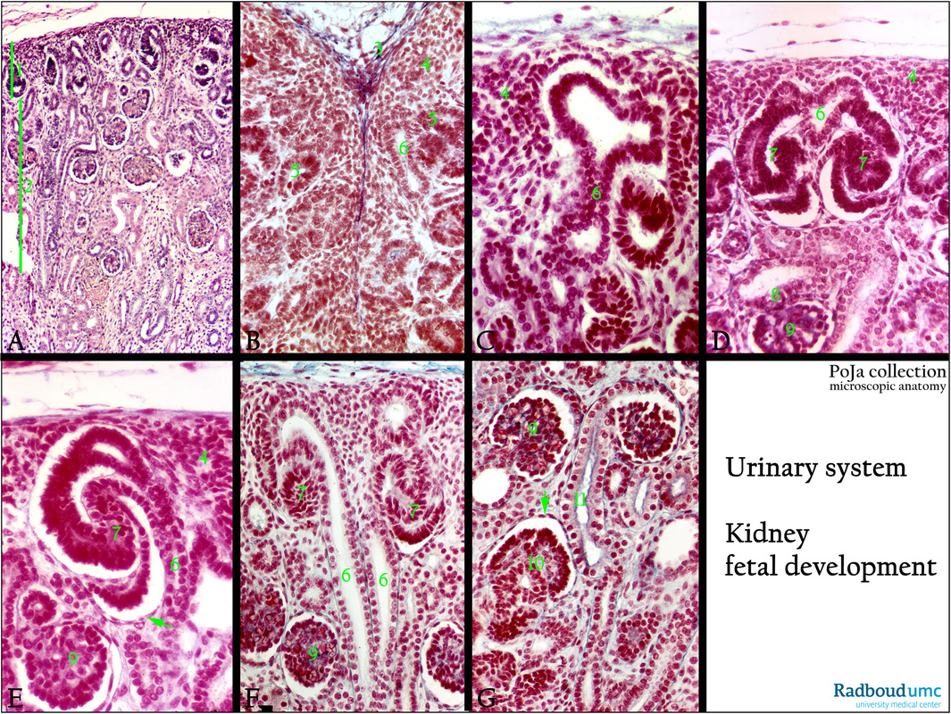
(A, B - G): Survey and details of kidney cortex, stain hematoxylin eosin in (A) and (B - G) stain Azan, human fetus.
(A): Shows the nephrogenic zone (bar 1) and below the differentiating nephrons (bar 2).
(B): Nephrogenic zone: Capsule (blue, 3) covering the cap mesenchyme (4) followed by renal vesicles (5), branch of ureteric tip (6).
(C): Cap mesenchyme (4). (6) Ureteric tip (later on collecting duct) with the T-shaped structure and its ampullae.
(D): T-structure of ureteric tip (6) with outgrowing ampullae (later on the distal tubules), S-shaped body (7), with the future visceral epithelial cells (podocytes) and opposite a thin future parietal lining, (6) collecting duct.
Note at the bottom immature glomerulus (9) close to future macula densa (8).
(E): Capsular lining (blue) with aggregation of metanephric mesenchyme (4). Ureteric tip (6) connected to part of the T-structure and
future distal tubule.
S-shaped body with future podocytes (7), thin future parietal lining (arrow). (9) nonmature glomerulus.
(F): Cap mesenchyme (4) around ureteric tip (6), S-shaped body (7), immature glomeruli (9).
(G): Immature glomeruli (9), future proximal duct (11) and late S-shaped body (10) with visceral epithelial cells separated by the future Bowman space from the thin parietal lining cells (arrow).
Background: 1) Metanephric nephron formation (in rodents):
-Under the influence of molecular markers a.o. Wnt9b, Pax8, Notch2 the following processes take place.
-Near the cloaca the nephric duct forms an outgrowth the uteric bud that grows into the metanephric mesenchyme
i.e. a population of specialized cells of the nephrogenic cord.
-Branching of the ureteric bud as well as formation of nephrons from the metanephric mesenchyme occur due to reciprocal inductive signalling between the bud and mesenchyme.
-Later on the ureteric bud branches and forms a T-shaped structure.
-Each bud is surrounded by a cap of condensed metanephric mesenchyme (cap mesenchyme).
-Cap mesenchyme cells form pretubular aggregates and the latter subsequently are converted into epithelial vesicles or renal vesicles
on either side of each ureteric bud tip.
-Growing renal vesicles result in the formation of comma- and S-shaped bodies during tubulogenesis.
-S-shaped bodies form most of the mature nephrons and fuse to ureteric bud branches which become future collecting ducts.
-Then blood vessel progenitors (angioblasts) invade the proximal cleft of the S-shaped body contributing to the vascular component of
the glomerulus.
-The distal part of the S-shaped body fuses with the collecting duct.
2) In humans nephrogenesis only occurs before birth but nephron maturation continues postnatally.
Generally there are four stages in human nephron development:
a) 13-19 weeks second trimester V-stage (Vesicle stage) is at its highest, after 20 weeks the V-stage proportion decreases gradually.
b) 20-24 weeks second trimester the proportion of the S-stage (S-shaped body stage) is at its maximum and epithelial and mesangial cells firstly appear during the stage.
c) 25-29 weeks third semester the C-stage (Capillary loop stage) proportion is high and maturation of endothelial cells starts.
d) 1-6 months infants, neonatal and postnatal M-stage (Maturation stage).
Keywords/Mesh: urinary system, embryo, fetus, kidney, cortex, nephrogenesis, glomerulus, histology, POJA collection
Title: Fetal kidney II
Description:
(A, B - G): Survey and details of kidney cortex, stain hematoxylin eosin in (A) and (B - G) stain Azan, human fetus.
(A): Shows the nephrogenic zone (bar 1) and below the differentiating nephrons (bar 2).
(B): Nephrogenic zone: Capsule (blue, 3) covering the cap mesenchyme (4) followed by renal vesicles (5), branch of ureteric tip (6).
(C): Cap mesenchyme (4). (6) Ureteric tip (later on collecting duct) with the T-shaped structure and its ampullae.
(D): T-structure of ureteric tip (6) with outgrowing ampullae (later on the distal tubules), S-shaped body (7), with the future visceral epithelial cells (podocytes) and opposite a thin future parietal lining, (6) collecting duct.
Note at the bottom immature glomerulus (9) close to future macula densa (8).
(E): Capsular lining (blue) with aggregation of metanephric mesenchyme (4). Ureteric tip (6) connected to part of the T-structure and
future distal tubule.
S-shaped body with future podocytes (7), thin future parietal lining (arrow). (9) nonmature glomerulus.
(F): Cap mesenchyme (4) around ureteric tip (6), S-shaped body (7), immature glomeruli (9).
(G): Immature glomeruli (9), future proximal duct (11) and late S-shaped body (10) with visceral epithelial cells separated by the future Bowman space from the thin parietal lining cells (arrow).
Background: 1) Metanephric nephron formation (in rodents):
-Under the influence of molecular markers a.o. Wnt9b, Pax8, Notch2 the following processes take place.
-Near the cloaca the nephric duct forms an outgrowth the uteric bud that grows into the metanephric mesenchyme
i.e. a population of specialized cells of the nephrogenic cord.
-Branching of the ureteric bud as well as formation of nephrons from the metanephric mesenchyme occur due to reciprocal inductive signalling between the bud and mesenchyme.
-Later on the ureteric bud branches and forms a T-shaped structure.
-Each bud is surrounded by a cap of condensed metanephric mesenchyme (cap mesenchyme).
-Cap mesenchyme cells form pretubular aggregates and the latter subsequently are converted into epithelial vesicles or renal vesicles
on either side of each ureteric bud tip.
-Growing renal vesicles result in the formation of comma- and S-shaped bodies during tubulogenesis.
-S-shaped bodies form most of the mature nephrons and fuse to ureteric bud branches which become future collecting ducts.
-Then blood vessel progenitors (angioblasts) invade the proximal cleft of the S-shaped body contributing to the vascular component of
the glomerulus.
-The distal part of the S-shaped body fuses with the collecting duct.
2) In humans nephrogenesis only occurs before birth but nephron maturation continues postnatally.
Generally there are four stages in human nephron development:
a) 13-19 weeks second trimester V-stage (Vesicle stage) is at its highest, after 20 weeks the V-stage proportion decreases gradually.
b) 20-24 weeks second trimester the proportion of the S-stage (S-shaped body stage) is at its maximum and epithelial and mesangial cells firstly appear during the stage.
c) 25-29 weeks third semester the C-stage (Capillary loop stage) proportion is high and maturation of endothelial cells starts.
d) 1-6 months infants, neonatal and postnatal M-stage (Maturation stage).
Keywords/Mesh: urinary system, embryo, fetus, kidney, cortex, nephrogenesis, glomerulus, histology, POJA collection