7.1 POJA-L1470+1474+1475
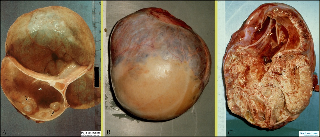
Title: Serous cystadenoma and serous adenocarcinoma of ovary (human, adult)
Description: (A, B, C) Macroscopy.
(A): Serous cystadenoma composed of fibrous cysts about 4-7 cm in diameter and lined by a smooth glinting surface wall. A few sessile papillae (arrows) are present.
(B): Expelled serous epithelial tumor in situ with smooth peritoneal covering. Nodular aspect is due to loculi.
(C): In cross-section it appears partly cystic and partly solid. The solid areas are formed of closely packed papillae. The cystic part (ca. 9 cm) contains blood-stained fluid. Due to large amounts of solid and papillary tumor mass this specimen is most probably a serous cystadenocarcinoma. (By courtesy of G. P. Vooijs MD, PhD, former Head of the Department of Pathology, and the Museum of Anatomy and Pathology, Radboud university medical center, Nijmegen, The Netherlands).
Background: It is assumed that the vast majority of serous and mucinous epithelial tumors of the ovary are derived from undifferentiated cells of the surface ovarian epithelium (serosa). The tumors arise either directly from that epithelium or from epithelial nests that are sequestrated into the cortex. Each type of neoplasm is presented as benign (adenoma) or as malignant (adenocarcinoma). The malignant tumors share common characteristics and are collectively known as ‘ovarian adenocarcinomas’.
Keywords/Mesh: female reproductive organs, ovary, adenocarcinoma, female genitalia, , ovarian neoplasms, serous cystadenoma, serous adenocarcinoma, pathology, macroscopy, histology, POJA collection

